Methods for large tissue labeling, clearing and imaging

A labeling and labeling agent technology, which is applied in the preparation of test samples, instruments, and analysis materials, etc., can solve the problems of imaging without single-cell resolution in adult mice, making tissues transparent, and analyzing the body, so as to reduce time and effort. Cost, Quantity Reduction, Conversion Boosting Effects

Pending Publication Date: 2020-04-10
DEEP PICTION GMBH
View PDF5 Cites 2 Cited by
  • Summary
  • Abstract
  • Description
  • Claims
  • Application Information

AI Technical Summary

Problems solved by technology

However, these methods do not actually analyze the whole body by fluorescence microscopy, only some anatomical parts of the body
Therefore, in these previous publications, there was no demonstration / evidence to image whole whole adult mice at single cell resolution
[0005] Also, until now, no technology has been able to make tissue as large as a pig brain transparent

Method used


Image

  • Methods for large tissue labeling, clearing and imaging
  • Methods for large tissue labeling, clearing and imaging
  • Methods for large tissue labeling, clearing and imaging

Examples


example 1

[0259] Example 1: Whole body immunolabeling of adult mice

[0260] The inventors set out to develop an immunostaining method for using fluorescent proteins to label cancer cells expressed throughout adult mice. The inventors reasoned that nanobodies are best suited to achieve immunolabelling of whole adult mice due to their small size (12-15 kDa compared to ~150 kDa for conventional antibodies) (Muyldermans, 2013; Yang et al., 2014). To deliver nanobodies conjugated to brightly colored Atto dyes (called nanoenhancers) throughout the body, the inventors developed a permeabilization solution containing Triton X-100, methyl-β-cyclodextrin (with for extracting cholesterol from biofilms) and trans-1-acetyl-4-hydroxy-L-proline (for loosening collagen networks) (Hama et al., 2015). The inventors circulated this permeabilization solution in mice (Gage et al., 2012; Ghanavati et al., 2014) via a high-pressure peristaltic pump (230mmHg, compared to 80-150mmHg for standard mouse heart...

example 2

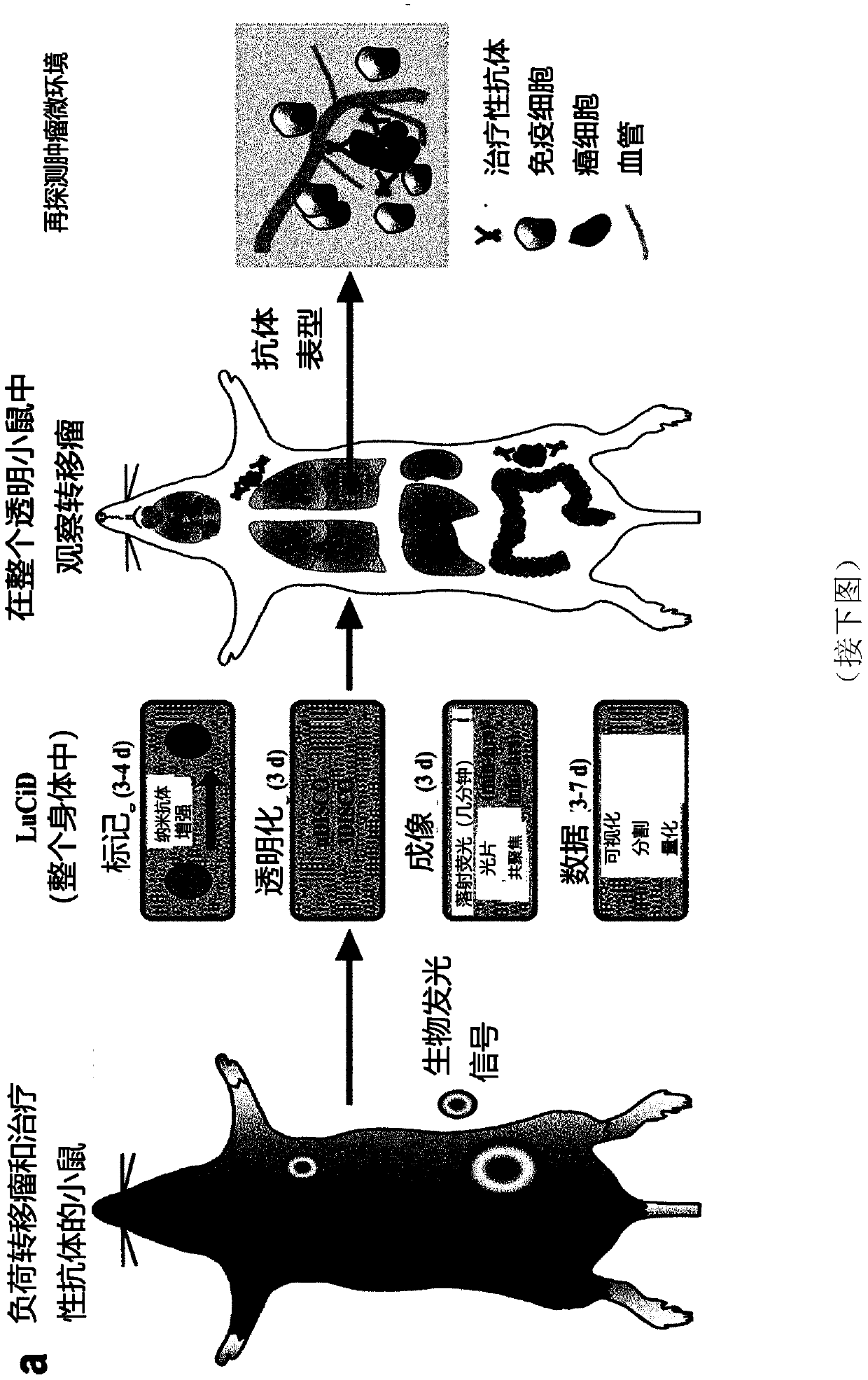
[0262] Example 2: Detection of tumor metastasis at single cell level in transparent mice

[0263] Many preclinical studies of cancer metastasis use mouse models using magnetic resonance imaging (MRI), computed tomography (CT), positron emission tomography (PET), single photon emission computed tomography (SPECT), or ultrasound and bioluminescent imaging (Condeelis and Weissleder, 2010; Massoud and Gambhir, 2003, 2007; Ntziachristos, 2010; Pichler et al., 2008; Timpson et al., 2011) and other methods to observe cancer cells at the primary site and in the distant body Regional growth. Although these methods provide important longitudinal information on the size of primary tumors and large metastases, they can usually only resolve structures larger than 50 μm (approximately 75 cells), so they are not capable of detecting smaller cells composed of fewer cells. The resolution of micrometastases. Because it is critical to detect smaller-sized clusters of tumor cells that may rep...

example 3

[0265] Example 3: Assessing therapeutic antibodies using LuCiD

[0266] Monoclonal antibodies targeting key tumor cell antigens are one of the most promising cancer treatments to emerge in the past two decades. A number of tumor-targeting monoclonal antibodies have become part of the standard of care for a variety of solid and hematologic malignancies, and many are in early or late stages of clinical development (Pandey and Mahadevan, 2014). Typically, therapeutic antibodies are raised against tumor-associated antigens overexpressed by cancer cells. Once these antibodies are injected, they are distributed throughout the body to target cancer cells. However, to date, there is no method for determining the distribution of therapeutic antibodies throughout the body at cellular resolution. Here, the inventors used LuCiD to assess the biodistribution of the therapeutic monoclonal antibody 6A10 against human CA12 (Battke et al., 2011). CA12 is overexpressed in various types of ...


PUM

PropertyMeasurementUnit
molecular weightaaaaaaaaaa

Abstract

The present invention relates to methods for preparing an animal tissue for fluorescence microscopy, to an animal tissue obtainable by said methods, to methods for analyzing said animal tissues, and to methods for the detection of metastases, for analyzing the biodistribution of a biopharmaceutical drug, and for analyzing the biodistribution of nanoparticles. The methods for preparing an animal tissue according to the present invention encompass whole-body labeling, clearing and imaging methods. The methods of the invention are advantageous in that they, for instance, allow the visualization of single cells within mammalian tissues including pig and human brains, tumor metastases at the single cell level and of the distribution of biopharmaceutical drugs (e.g. the distribution of cancer-targeting therapeutic antibodies in whole animals such as intact mice) at single cell level in whole mouse.

Description

technical field [0001] The present invention relates to methods for preparing animal tissues for fluorescence microscopy, animal tissues obtained according to said methods, methods for analyzing said animal tissues, as well as methods for detecting metastases, analyzing biodistribution of biopharmaceutical drugs and analyzing nanoparticles Methods of biodistribution. Methods of preparing animal tissue according to the invention include whole body labeling, clearing and imaging methods. The methods of the present invention are advantageous because they allow, for example, single-cell, metastases at the single-cell level within mammalian tissues, including pig and human brain, and biopharmaceutical drug distribution at the single-cell level throughout mice. (eg, visualization of the distribution of cancer-targeting therapeutic antibodies in a whole animal, such as a whole mouse). Background technique [0002] Understanding the details of the cells at which tumors invade and ...

Claims


Application Information

Patent Timeline
Patent Type & Authority Applications(China)
IPC IPC(8): G01N1/30G01N33/574
CPCG01N1/30G01N33/57492G01N33/582G01N33/6896G01N2800/28G01N1/286G01N21/6458G01N33/5005G01N33/5088G01N33/57496G01N33/6893G01N2001/2873G01N2001/302
Inventor 阿里·埃蒂尔克
Owner DEEP PICTION GMBH
Why Eureka
Try Eureka