Static artery separation method and device based on CT image

A CT image and separation method technology, applied in image analysis, image enhancement, image data processing, etc., can solve the problems of difficult standardization, time-consuming, unsuitable for large-scale clinical research, etc., achieve the effect of precise vein and artery separation and improve efficiency

Pending Publication Date: 2020-04-17
杭州健培科技有限公司
View PDF5 Cites 6 Cited by
  • Summary
  • Abstract
  • Description
  • Claims
  • Application Information

AI Technical Summary

Problems solved by technology

This process is time-consuming and difficult to standardize, making it unsuit

Method used


Image

  • Static artery separation method and device based on CT image
  • Static artery separation method and device based on CT image
  • Static artery separation method and device based on CT image

Examples


Embodiment Construction

[0037] In order to make the purpose, technical solutions and advantages of the present invention clearer, the present invention will be further described in detail below in conjunction with the accompanying drawings. Obviously, the described embodiments are only some of the embodiments of the present invention, rather than all of them.

[0038] Based on the embodiments of the present invention, all other embodiments obtained by persons of ordinary skill in the art without making creative efforts belong to the protection scope of the present invention.

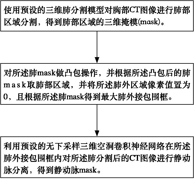
[0039] figure 1 A flowchart corresponding to a method for separating veins and arteries based on CT images provided by the present invention is shown. The flowchart can be executed by a device for separating veins and arteries based on CT images, including the following steps:

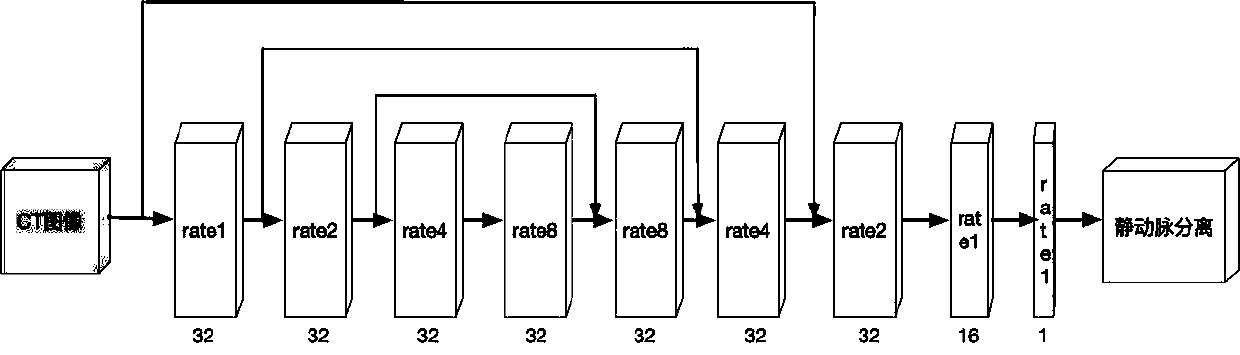
[0040] Step 101 , using a preset three-dimensional lung segmentation model to perform lung region segmentation on a chest CT image to obtain a three-di...


PUM


Abstract

The invention discloses a static artery separation method and device based on a CT image, and the method and device improve the efficiency and precision of static artery separation compared with a conventional method and manual labeling of a doctor, achieve the full-automatic static artery separation, and do not need the manual intervention. The method mainly comprises the following steps: carrying out lung region segmentation on a chest CT image by using a preset three-dimensional lung segmentation model to obtain a three-dimensional mask of a lung region; carrying out convex hull operation on a lung mask, taking a lung region according to the lung mask subjected to convex hull operation, setting the pixel value of the extrapulmonary region to be 0, and obtaining a maximum extrapulmonarybounding box according to the lung mask; and performing static artery separation on the segmented CT image of the lung in the lung external bounding box by using a preset downsampling-free three-dimensional cavity convolutional neural network to obtain a static artery mask. As the accurate annotation data is used for training, and the three-dimensional cavity convolutional neural network is used for learning, the loss of information amount is reduced, and the accurate separation of the static artery is realized.

Description

technical field [0001] The invention relates to the field of medical image processing, in particular to a method and device for separating veins and arteries based on CT images. Background technique [0002] Over the past few decades, computed tomography (CT) has become the most common imaging modality for diagnosing and evaluating lung disease. Modern CT scanners combined with modern imaging techniques allow (semi)automatic identification and extraction of lung structures, such as blood vessels and bronchi, with high accuracy using low radiation doses. However, although some progress has been made in CT image segmentation techniques in recent years, there are still many problems that have not been resolved. Among them, the identification and differentiation of pulmonary arteries and pulmonary veins is one of the most challenging problems. [0003] Dividing the pulmonary vessels into arteries / veins (A / Vs) may help doctors accurately diagnose lung diseases that may affect t...

Claims


Application Information

Patent Timeline
IPC IPC(8): G06T7/11G06T7/194
CPCG06T7/11G06T7/194G06T2207/10081G06T2207/20081G06T2207/20084G06T2207/30101G06T2207/30061
Inventor 姜志强程国华何林阳季红丽
Owner 杭州健培科技有限公司
Why Eureka
Try Eureka