Neural fiber extraction method, system and device for region of interest and storage medium
A region of interest and nerve fiber technology, applied in the field of medical image processing, can solve the problems of low efficiency and complicated process of nerve fiber tracking, and achieve the effect of improving tracking efficiency
- Summary
- Abstract
- Description
- Claims
- Application Information
AI Technical Summary
Problems solved by technology
Method used
Image
Examples
Embodiment 1
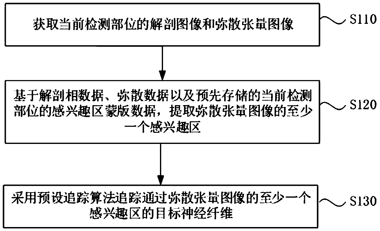
[0027] figure 1 It is a flow chart of a method for extracting nerve fibers in a region of interest provided by Embodiment 1 of the present invention. This embodiment is applicable to automatically extracting a region of interest and performing nerve fiber tracking based on the automatically extracted region of interest. The method can be implemented by a system for extracting nerve fibers in a region of interest, wherein the device can be realized by software and / or hardware, and generally integrated in a device for extracting nerve fibers in a region of interest. Specifically include the following steps:
[0028] S110. Acquire the anatomical image and the diffusion tensor image of the currently detected part.
[0029] Wherein, the current detection site may be the head, the chest, or other sites. Optionally, the head is used as the current detection site for introduction in this embodiment.
[0030] Diffusion tensor image is a kind of magnetic resonance image obtained by co...
Embodiment 2
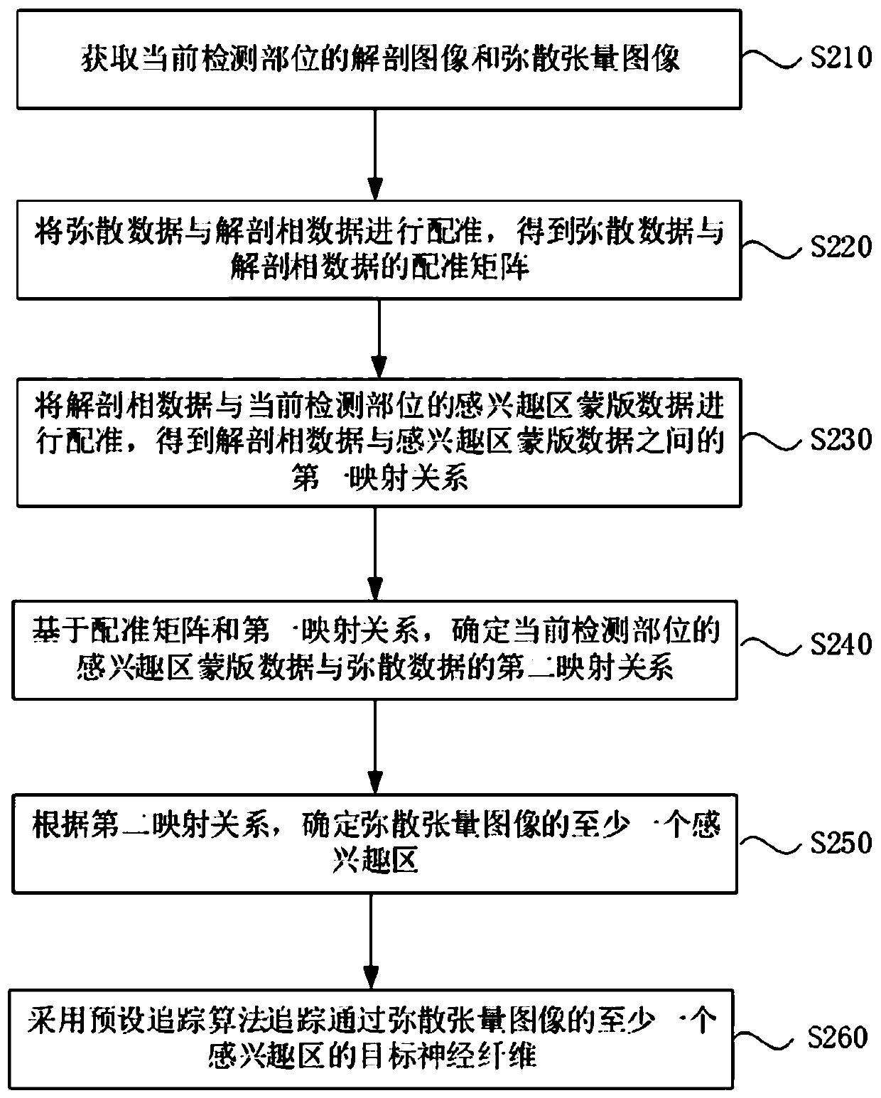
[0042] figure 2It is a schematic flowchart of a method for extracting nerve fibers in a region of interest provided in Embodiment 2 of the present invention. The technical solution of this embodiment is refined on the basis of the above embodiments. Optionally, the ROI mask based on the anatomical phase data, the diffusion data, and the pre-stored current detection part data, extracting at least one region of interest of the diffusion tensor image, comprising: registering the diffusion data and the anatomical phase data to obtain a registration matrix of the diffusion data and the anatomical phase data; Registering the anatomical phase data with the ROI mask data of the current detected part to obtain a first mapping relationship between the anatomical phase data and the ROI mask data; based on the registration Matrix and the first mapping relationship, determine the second mapping relationship between the mask data of the region of interest of the current detected part and ...
Embodiment 3
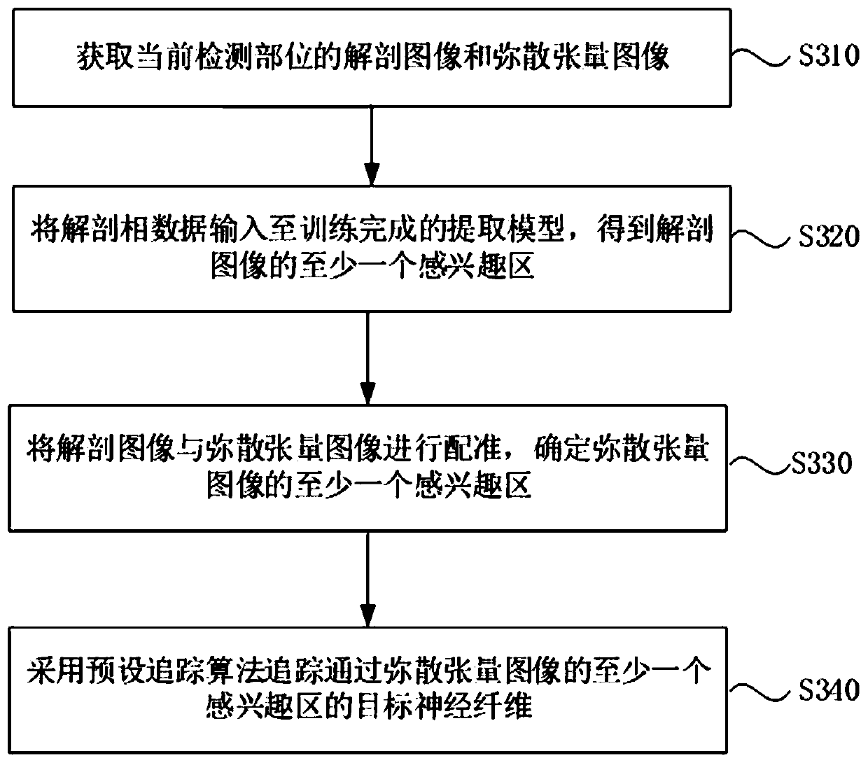
[0056] image 3 It is a schematic flowchart of a method for extracting nerve fibers in a region of interest provided in Embodiment 3 of the present invention. The technical solution of this embodiment extracts at least one region of interest of the diffusion tensor image based on deep learning. Optionally, extracting at least one region of interest of the diffusion tensor image includes: inputting the anatomical phase data into The trained second extraction model obtains at least one region of interest of the anatomical image; wherein, the second extraction model obtains the region of interest of the multiple historical anatomical images and the anatomy of the multiple historical anatomical images obtained by manually dividing The phase data is obtained by training the second original neural network model; the anatomical image is registered with the diffusion tensor image, and at least one region of interest of the diffusion tensor image is determined. For details, see imag...
PUM
 Abstract
Description
Claims
Application Information
 - R&D
- Intellectual Property
- Life Sciences
- Materials
- Tech Scout
- Unparalleled Data Quality
- Higher Quality Content
- 60% Fewer Hallucinations
Browse by: Latest US Patents, China's latest patents, Technical Efficacy Thesaurus, Application Domain, Technology Topic, Popular Technical Reports.
© 2025 PatSnap. All rights reserved.Legal|Privacy policy|Modern Slavery Act Transparency Statement|Sitemap|About US| Contact US: help@patsnap.com


