Method for detecting NSPs residues in foot-and-mouth disease inactivated antigens or inactivated vaccines

A technology of inactivated vaccines and inactivated antigens, applied in the field of immunology, can solve the problems of expensive, time-consuming, inability to determine which kinds of NSPs antigens or vaccines have, and achieve the effects of strong specificity, high sensitivity and simple operation.

Inactive Publication Date: 2020-06-05
LANZHOU INST OF VETERINARY SCI CHINESE ACAD OF AGRI SCI
View PDF5 Cites 1 Cited by
  • Summary
  • Abstract
  • Description
  • Claims
  • Application Information

AI Technical Summary

Problems solved by technology

However, animal experiments are expensive and time-consuming. If a batch of vaccines cannot meet the purity requirements, the manufacturer must discard the entire batch of vaccines.
[0003] At present, there is no method f

Method used


Image

  • Method for detecting NSPs residues in foot-and-mouth disease inactivated antigens or inactivated vaccines
  • Method for detecting NSPs residues in foot-and-mouth disease inactivated antigens or inactivated vaccines
  • Method for detecting NSPs residues in foot-and-mouth disease inactivated antigens or inactivated vaccines

Examples


Embodiment 1

[0039] Example 1 Reactivity of 3B monoclonal antibody with FMDV O / NC / CHA / 2010 infected cells

[0040] 1. Preparation of 3B monoclonal antibody

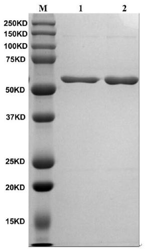
[0041] The foot-and-mouth disease NSP 3ABC of prokaryotic expression is purified ( figure 1 ), and after gradient dialysis and renaturation, the mice were immunized to prepare monoclonal antibodies, the positive hybridoma cells were subcloned three times by the limiting dilution method, and the highly reactive monoclonal cells were screened out, and then the screened cells were injected into small The ascites fluid was prepared from the peritoneal cavity of the rat and purified, and the purified monoclonal antibody was coupled with horseradish peroxidase (HRP) (9E2-HRP). Verification by Western blot showed that the monoclonal antibody recognized 3B protein ( figure 2 ).

[0042] 2. Expression of NSPs in BHK-21 infected with O-type FMDV at different times

[0043] Inoculation: Resuspend the BHK-21 digested with trypsin and add it ...

Embodiment 2

[0049] Embodiment 2 detects NSPs in the foot-and-mouth disease inactivated antigen

[0050] Take 60 μl of FMD antigen and add 20 μl of 4× loading buffer, then place it in boiling water for 10 min. Add the boiled samples and markers into protein gel wells, electrophoresis, transfer to membrane, block, react with 9E2-HRP, expose or scan the membrane (the specific steps are the same as in Example 1).

[0051] Depend on Figure 4 It can be known that when detecting NSPs residues in unpurified antigen (0#), once-purified antigen (1#) and twice-purified antigen (2#), the unpurified and once-purified antigens were both 3ABB, 3ABBB and 3ABC degradation products (3ABC-) were detected, while the antigen purified twice was not detected. Compared with unpurified antigens, NSPs residues in antigens purified once have been greatly reduced. When detecting concentrated antigens (1-10), it can be observed that 3ABB, 3ABBB and 3ABC- are detected for all antigens 1-10, while there are no band...

Embodiment 3

[0053] Embodiment 3 detects NSPs in the finished product vaccine of foot-and-mouth disease

[0054] Take 1.5 mL of the vaccine and place it in a 2 mL centrifuge tube, then add 75 μL of n-pentanol to vortex for 30 s, and then stand at 4°C for 1 h. After complete demulsification, centrifuge at 8,000 g for 10 min, absorb the lower aqueous phase, centrifuge at 8,000 g for 10 min, take 60 μl, then add 20 μl of 4× loading buffer, and boil it in boiling water for 10 min. Add the cooked samples and markers to the protein gel wells, perform electrophoresis, transfer to membrane, block, react with 9E2-HRP, expose or scan the membrane (the specific steps are the same as in Example 1).

[0055] Depend on Image 6 It can be seen that when testing the finished vaccines A-L, NSPs 3ABBB and 3ABC- were only detected in C, F and K, and the amount detected in K was large, and NSPs 3ABB was also detected.


PUM


Abstract

The invention relates to a method for detecting NSPs residues in foot-and-mouth disease inactivated antigens or inactivated vaccines, and belongs to the technical field of immunology. The method comprises the following steps: carrying out protein electrophoresis on an inactivated antigen and a demulsified foot-and-mouth disease inactivated vaccine so as to separate proteins with different sizes inthe antigen or the vaccine, then carrying out WB, and judging whether the antigen or the vaccine contains NSPs and components of the NSPs according to an appeared band and standard contrast. The method has the advantages of strong specificity, high sensitivity and simple operation, can be operated in a common laboratory, is very suitable for foot-and-mouth disease manufacturers, and provides technical guidance for the foot-and-mouth disease manufacturers to purify antigens and formulate standardized processes.

Description

technical field [0001] The invention belongs to the technical field of immunology, and in particular relates to a method for detecting NSPs residues in inactivated antigens of foot-and-mouth disease or inactivated vaccines. Background technique [0002] Foot-and-mouth disease (FMD) is caused by foot-and-mouth disease virus (FMDV), an acute, febrile, highly contagious animal disease that infects pigs, cattle, sheep and other cloven-hoofed animals. FMDV belongs to the genus Foot-and-Mouth Disease Virus of the family Picornaviridae, encoding 4 structural proteins (SPs: VP4, VP2, VP3 and VP1) and 10 non-structural proteins (NSPs: L, 2A, 2B, 2C, 3A, 3B, 3C, 3D , 3AB, 3ABC). The prevention and control of foot-and-mouth disease in my country mainly adopts comprehensive prevention and control measures of vaccine immunization, culling of infected and suspicious animals, and serum monitoring to control the prevalence of foot-and-mouth disease. Among them, inactivated vaccines are mai...

Claims


Application Information

Patent Timeline
IPC IPC(8): G01N33/569G01N33/577G01N33/58
CPCG01N33/56983G01N33/577G01N33/581G01N2333/09G01N2469/10
Inventor 常惠芸刘伟邵军军
Owner LANZHOU INST OF VETERINARY SCI CHINESE ACAD OF AGRI SCI
Why Eureka
Try Eureka