Survival myocardial evaluation method

An evaluation method and myocardial technology, applied in the field of medical image diagnosis, can solve problems such as unestablished, and achieve the effects of good consistency, high repeatability, and good diagnostic accuracy

Active Publication Date: 2020-06-19
THE FIRST PEOPLES HOSPITAL OF CHANGZHOU
View PDF4 Cites 2 Cited by
  • Summary
  • Abstract
  • Description
  • Claims
  • Application Information

AI Technical Summary

Problems solved by technology

However, the method of evaluating myocardial viability using gated SPECT myocardial

Method used


Image

  • Survival myocardial evaluation method
  • Survival myocardial evaluation method
  • Survival myocardial evaluation method

Examples


Embodiment 1

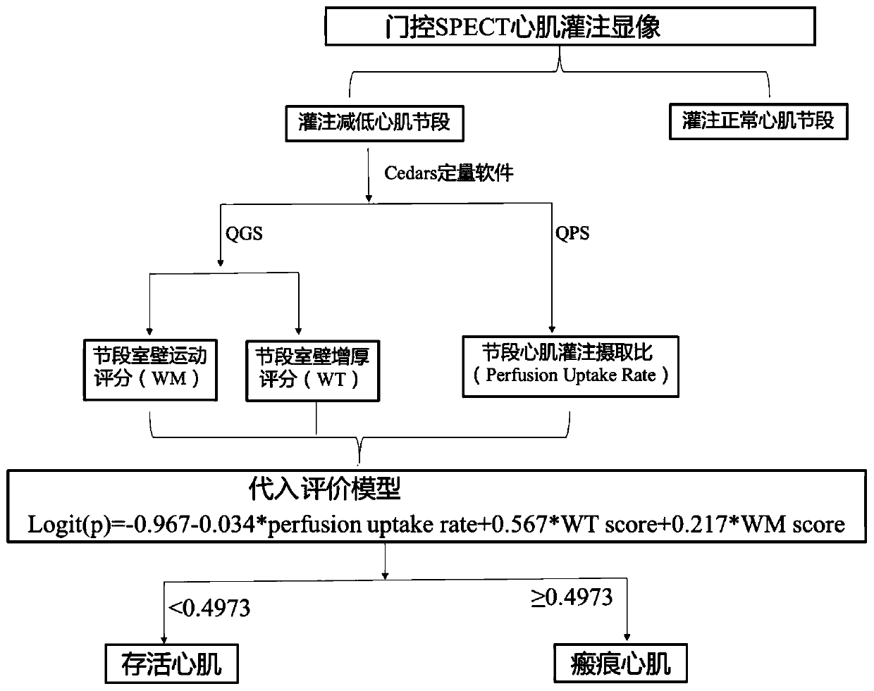
[0029] like figure 1 As shown, Example 1 discloses a method for evaluating viable myocardium, which is based on gated SPECT myocardial perfusion imaging for evaluating myocardial function combined with myocardial perfusion, specifically including the following steps:

[0030] Step 1: Use gated SPECT myocardial perfusion imaging to obtain inspection images, and reconstruct left ventricular short-axis, horizontal long-axis, and vertical long-axis images; in this embodiment 1, gated SPECT myocardial perfusion imaging is 99m Tc-MIBI resting gated SPECT myocardial perfusion imaging was implemented using a dual-probe SPECT / CT instrument (Symbia T16, Siemens Medical Systems, Erlangen, Germany) from Siemens, Germany, and the imaging agent was provided by Shanghai Xinke Pharmaceutical Co., Ltd. of 99m Tc-MIBI, radiochemical purity >95%, injection dose 740-925MBq; acquisition conditions: parallel hole low-energy high-resolution collimator, energy peak 140keV, window width 20%, matrix 1...

Embodiment 2

[0038] Concrete process and effectiveness of the present invention are specified below with concrete example, specifically as follows:

[0039] 1. Research purpose: To analyze the gain value of gated SPECT myocardial perfusion imaging quantitative analysis of myocardial function in evaluating the viability of myocardium in patients with coronary heart disease and heart failure, and to obtain a method for evaluating viable myocardium based on gated SPECT myocardial perfusion imaging.

[0040] 2. Research design: The clinical trial method of single-center cross-sectional retrospective study was adopted.

[0041] 3. Case selection:

[0042] 3.1. Inclusion criteria:

[0043] 1) Myocardial infarction over 3 months confirmed by medical history, echocardiography and electrocardiogram (ECG); 2) Coronary heart disease patients with single or multiple vessel stenosis of more than 50% diagnosed by coronary angiography (CAG); 3 ) Severe left ventricular dysfunction, defined as left vent...


PUM


Abstract

The invention relates to a survival myocardial evaluation method, which is used for evaluating based on gating SPECT myocardial perfusion development myocardial functions in combination with myocardial perfusion, and comprises the following steps: firstly, acquiring an examination image by adopting gating SPECT myocardial perfusion development, and reconstructing to obtain a left ventricular minoraxis image, a left ventricular horizontal major axis image and a left ventricular vertical major axis image; then carrying out semi-quantitative scoring on blood perfusion uptake of the left ventricular myocardial segment, and judging a perfusion reduction segment; quantitatively evaluating to obtain the blood perfusion uptake ratio of each segment of the left ventricular myocardium; analyzing the reconstructed gated SPECT myocardial perfusion imaging, respectively obtaining a ventricular wall movement score and a thickening rate score of each myocardial segment, and finally substituting theparameters into an evaluation model for calculation. Evaluation is carried out based on gated SPECT myocardial perfusion imaging myocardial functions in combination with myocardial perfusion, and themethod is particularly suitable for evaluating survival myocardium of coronary heart disease heart failure patients before blood supply reconstruction and guiding clinical treatment decisions.

Description

technical field [0001] The invention relates to an evaluation method of intermediate clinical indications, in particular to an evaluation method of viable myocardium, which belongs to the technical field of medical image diagnosis. Background technique [0002] Viable myocardium refers to ischemic myocardium with dysfunction, and its function can gradually recover after blood perfusion is restored. Revascularization therapy can improve the cardiac function and prognosis of patients with coronary heart disease by restoring the blood perfusion of ischemic myocardium, and is an important means of treating coronary heart disease. At present, the number of viable myocardium has been widely used to predict the recovery of systolic function after revascularization in patients with coronary heart disease and heart failure. The reversal of ventricular remodeling, the improvement of quality of life, exercise capacity and survival rate are important basis for guiding clinical decision-...

Claims


Application Information

Patent Timeline
IPC IPC(8): G06T7/00G16H50/20A61K51/04A61K101/02A61K103/10
CPCG06T7/0012G16H50/20A61K51/0491A61K51/0478G06T2207/10081G06T2207/30101G06T2207/30048Y02A90/10
Inventor 王跃涛张飞飞王建锋邵晓梁孙永红邱春
Owner THE FIRST PEOPLES HOSPITAL OF CHANGZHOU
Why Eureka
Try Eureka