Exosome separation and purification device and separation and purification method thereof

A technology for separation and purification of exosomes, applied in the field of medical experiment analysis, can solve the problems of high cost, large loss of sample volume, failure of filtration and processing methods, etc.

Pending Publication Date: 2020-07-07
苏州先觉生物科技有限公司
View PDF0 Cites 3 Cited by
  • Summary
  • Abstract
  • Description
  • Claims
  • Application Information

AI Technical Summary

Problems solved by technology

[0004] Ultracentrifugation is currently the "gold standard" of exosome isolation methods. However, ultracentrifugation technology has the disadvantages of time-consuming, expensive, and large sample loss. Membrane filtration technology can separate and purify exosomes. When the sample size Larger use of filtration membranes to separate and purify exosome vesicles is easy to block or block, and whether exosomes pass through the filtration membrane is easy to adhere and cannot be eluted
[0005] In the prior art, the Chinese invention patent No. ZL2016105973232, titled "Extraction Method of Exosomes in Human Body Fluids" discloses "a. Preparation of body fluid samples: first extract human body fluids under sterile conditions and dilute them with PBS buffer. Then preliminarily remove the cell components and cell fragments in the body fluid by centrifugal screening, and make a body fluid sam...

Method used


Image

  • Exosome separation and purification device and separation and purification method thereof
  • Exosome separation and purification device and separation and purification method thereof
  • Exosome separation and purification device and separation and purification method thereof

Examples


Embodiment 1

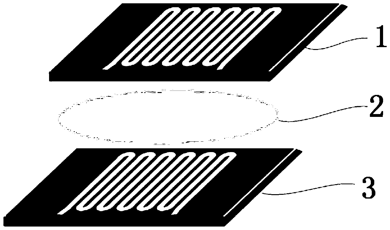
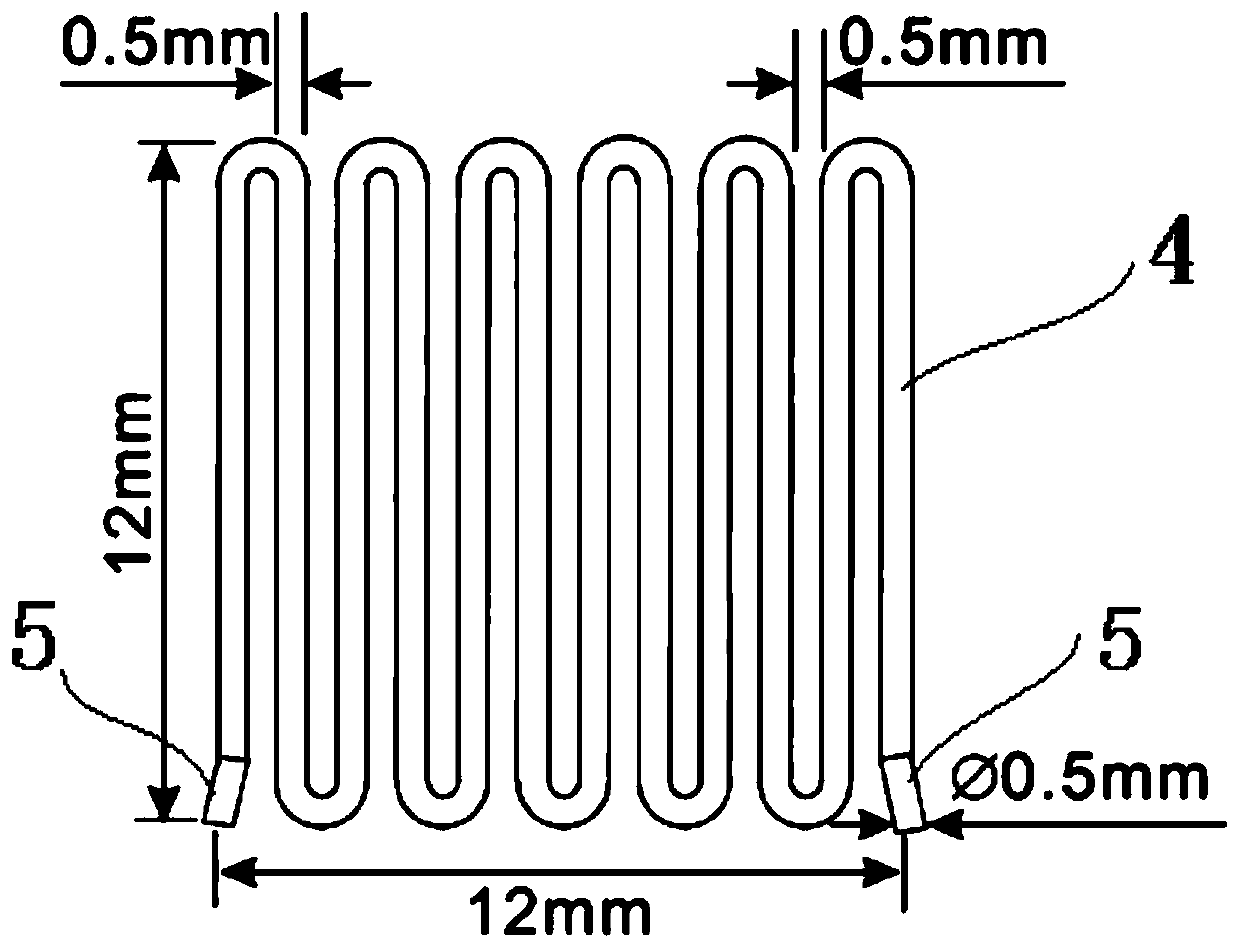
[0035] Such as figure 1 The shown exosome separation and purification device of the present invention includes an upper microfluidic chip 1, a filter membrane 2 and a lower microfluidic chip 3. The microfluidic chip is an existing technology in the field of medical research and is mainly used for For accurate input of quantitative detection liquid, the filter membrane 2 is arranged between the upper microfluidic chip 1 and the lower microfluidic chip 3, such as figure 1 As shown, the present invention adopts the three-layer setting of the chip, the filter membrane 2 and the chip. The improved setting greatly reduces the demand for sample liquid for exosome separation and purification, and the filtering effect is more controllable and accurate than direct filtering. The upper microfluidic chip 1 and the lower microfluidic chip 3 are respectively provided with two inlets 5, and the sample liquid is injected inward through one of the inlets 5 of the upper microfluidic chip 1, and...

Embodiment 2

[0043] A separation and purification method using the exosome separation and purification device described in Example 1, comprising the following steps:

[0044] (1) One of the inlets 5 of the upper microfluidic chip 1 and the lower microfluidic chip 3 is blocked respectively, and the two blocked inlets 5 are two ports on the diagonal, so that the liquid sample to be tested is in the In the farthest distance of the flow in the microfluidic chip, the unimpeded inlet 5 of the upward microfluidic chip 1 injects the liquid sample to be tested into the upper microfluidic chip 1 at a flow rate of 10ul / min;

[0045] (2) After the liquid sample to be tested passes through the upper microfluidic chip 1, it enters the filter membrane 2 for filtration to remove pollutants and enrich exosomes. outflow in 5;

[0046] (3) In the unimpeded inlet 5 of the upward microfluidic chip 1, air is introduced at a flow rate of 100ul / min to completely remove the liquid in the channel;

[0047] (4) In...

Embodiment 3

[0054] In the exosome separation and purification device and its separation and purification method described in Examples 1 and 2, the following specific experiments were carried out:

[0055] 2.1 Equipment source:

[0056] Ultracentrifuge: Optima XE-90Ultracentrifuge (BECKMAN COULTER);

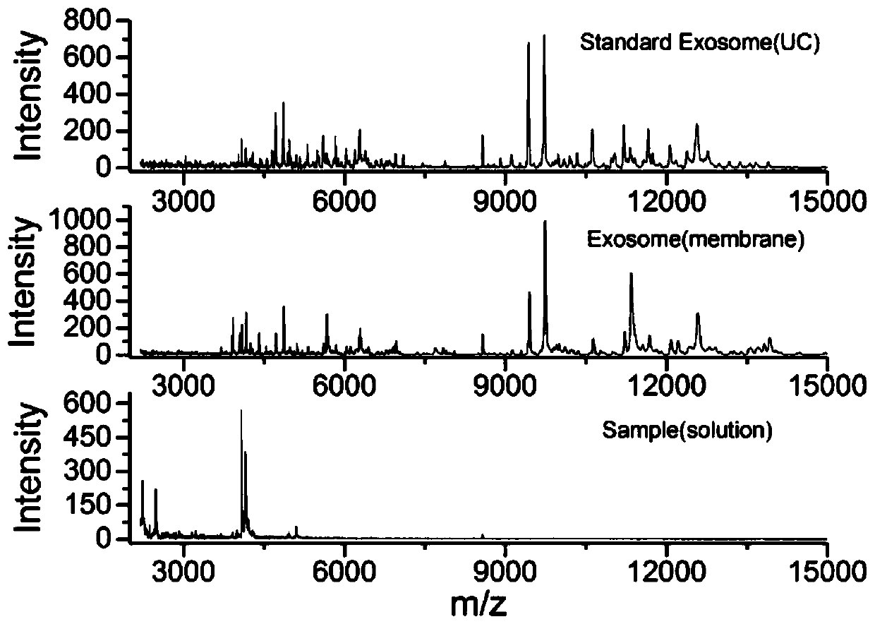
[0057] MALDI-TOF mass spectrometer: Bruker Microflex LRF MALDI-TOF mass spectrometer (Bremen, Germany).

[0058] Matrix-assisted laser desorption ionization time-of-flight mass spectrometry (Matrix-assisted laserdesorption / ionization time-of-flight mass spectrometry, MALDI-TOF MS) has been developed since the 1980s, and has ultra-high sensitivity and specificity. The molecular weight of biomolecules can be accurately determined to realize the identification and analysis of biomacromolecules; by analyzing the fingerprints of different bacteria, MALDI-TOF MS can realize the rapid identification of medical experiments; in addition, MALDI-TOF MS technology is also widely used in proteomics resea...


PUM

PropertyMeasurementUnit
Widthaaaaaaaaaa
Depthaaaaaaaaaa
Apertureaaaaaaaaaa

Abstract

The invention relates to an exosome separation and purification device and a separation and purification method thereof. The device comprises an upper micro-fluidic chip, a filter membrane and a lowermicro-fluidic chip, and the filter membrane is arranged between the upper micro-fluidic chip and the lower micro-fluidic chip. Sample liquid is injected into the upper micro-fluidic chip through oneinlet of the upper micro-fluidic chip, and after the sample liquid is filtered through the filtering membrane, an exosome is obtained from one inlet of the lower micro-fluidic chip. According to the method disclosed by the invention, impurity protein in the exosome can be removed, the whole exosome detection and analysis time of the technology is less than 1 hour, and the technology is stable. A sample obtained by the device can be directly subjected to mass spectrometry, and the main signals of a graph obtained are consistent with those of a mass spectrum detection graph obtained after exosome cyst separation and purification through an existing standard ultracentrifugation method. The method has the advantages of being low in cost, small in amount of samples needed, rapid, convenient touse, accurate, stable, capable of avoiding the defects that time and resources are consumed in the ultracentrifugation technology and the like and wide in biological and medical application prospect.

Description

technical field [0001] The invention relates to an exosome separation and purification device and a separation and purification method thereof, belonging to medical experiment analysis methods. Background technique [0002] Exosomes are a kind of extracellular vesicles (Extracellular Vesicles, EV, the size of extracellular vesicles is about 30-1000nm), and its diameter is about 30-150nm. Rich in membrane proteins. Exosome vesicles contain proteins, nucleic acids (miRNA, mRNA, etc.), membrane proteins, or lipids. [0003] Microfluidics integrates the basic operation units such as sample preparation, reaction, separation, and detection in the biological, chemical, and medical analysis processes into a micron-scale chip to automatically complete the entire analysis process. Due to its great potential in the fields of biology, chemistry, and medicine, it has developed into a new research field interdisciplinary in biology, chemistry, medicine, fluid, electronics, materials, an...

Claims


Application Information

Patent Timeline
IPC IPC(8): G01N1/34G01N27/64B01L3/00
CPCB01L3/5027G01N1/34G01N27/64
Inventor 沈宇辉
Owner 苏州先觉生物科技有限公司
Why Eureka
Try Eureka