Model generation method, medical image segmentation method and device, equipment and medium

A model generation and image segmentation technology, which is applied in the field of medical image processing, can solve the problems that the segmentation accuracy of the lung area needs to be improved, and the unique characteristics of the lung area are not considered, so as to achieve the effect of strong generalization performance and improved matching degree

Active Publication Date: 2020-07-17
INFERVISION MEDICAL TECH CO LTD
View PDF5 Cites 7 Cited by
  • Summary
  • Abstract
  • Description
  • Claims
  • Application Information

AI Technical Summary

Problems solved by technology

However, these network models do not take into account the unique characteristics of the lung region when they are set up, and they use t

Method used


Image

  • Model generation method, medical image segmentation method and device, equipment and medium
  • Model generation method, medical image segmentation method and device, equipment and medium
  • Model generation method, medical image segmentation method and device, equipment and medium

Examples


Embodiment 1

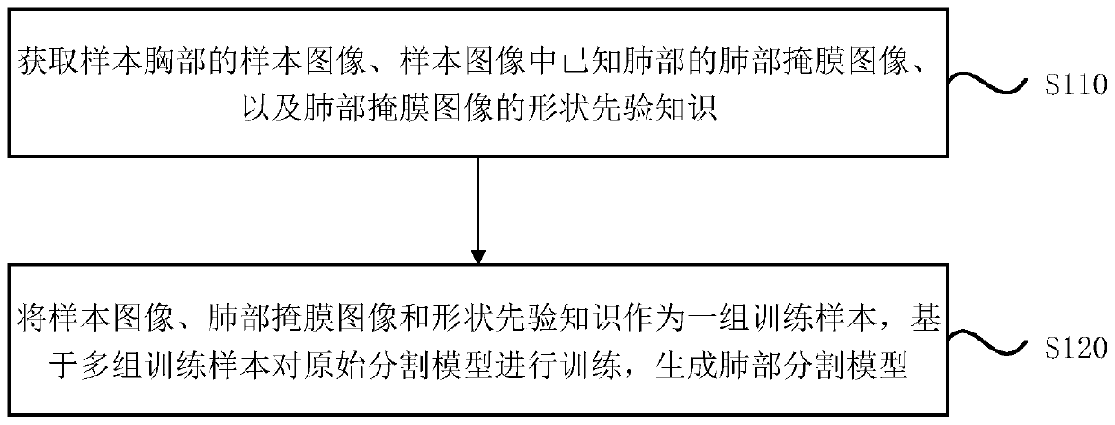
[0049] figure 1 It is a flowchart of a model generation method provided in Embodiment 1 of the present invention. This embodiment is applicable to the case of generating a lung segmentation model matching the lung region segmentation, especially applicable to the case of generating a lung segmentation model matching the lung region segmentation based on a multi-task learning framework. The method can be executed by the model generation device provided by the embodiment of the present invention, the device can be realized by software and / or hardware, and the device can be integrated on various user terminals or servers.

[0050] see figure 1 , the method of the embodiment of the present invention specifically includes the following steps:

[0051] S110. Acquire a sample image of the sample chest, a lung mask image of known lungs in the sample image, and shape prior knowledge of the lung mask image.

[0052]Wherein, the sample image can be obtained based on X-ray imaging tech...

Embodiment 2

[0071] Figure 5 It is a flowchart of a medical image segmentation method provided in Embodiment 2 of the present invention. This embodiment is applicable to the case of segmenting the lung area in the X-ray chest film, especially suitable for the case of segmenting the lung area in the X-ray chest film based on the lung segmentation model, the lung segmentation model is in The prior knowledge of the shape of the lung region is effectively utilized during the training process. The method can be executed by the medical image segmentation device provided by the embodiment of the present invention, the device can be realized by software and / or hardware, and the device can be integrated on various user terminals or servers.

[0072] see Figure 5 , the method of the embodiment of the present invention specifically includes the following steps:

[0073] S210. Acquire a subject image of the subject chest and a trained lung segmentation model generated according to the model gener...

Embodiment 3

[0078] Figure 6 It is a structural block diagram of a model generation device provided in Embodiment 3 of the present invention, and the device is used to execute the model generation method provided in any of the above embodiments. The device and the model generation method in the above-mentioned embodiments belong to the same inventive concept. For details not described in detail in the embodiments of the model generation device, reference may be made to the above-mentioned embodiment of the model generation method. see Figure 6 , the device may specifically include: a first acquisition module 310 and a model generation module 320 .

[0079] Wherein, the first acquisition module 310 is used to acquire the sample image of the sample chest, the lung mask image of known lungs in the sample image, and the prior knowledge of the shape of the lung mask image;

[0080] The model generation module 320 is used to use the sample image, the lung mask image and shape prior knowledge...


PUM


Abstract

The embodiment of the invention discloses a model generation method, a medical image segmentation method and device, equipment and a medium. The model generation method comprises the steps of obtaining a sample image of a sample chest, a lung mask image of a known lung in the sample image, and shape priori knowledge of the lung mask image; taking the sample image, the lung mask image and the shapepriori knowledge as a group of training samples, and training the original segmentation model based on multiple groups of training samples to generate a lung segmentation model, wherein the originalsegmentation model comprises a feature extraction network, and an image segmentation network and a priori knowledge regression network which are respectively connected with the feature extraction network. According to the technical scheme of the embodiment of the invention, the problem that the existing segmentation model does not effectively utilize the shape priori knowledge of the lung region is solved, and the priori knowledge regression network set based on the shape priori knowledge is matched with the existing image segmentation network, so that the matching degree of the lung segmentation model and lung region segmentation is improved.

Description

technical field [0001] Embodiments of the present invention relate to the technical field of medical image processing, and in particular to a model generation method, a medical image segmentation method, a device, a device, and a medium. Background technique [0002] X-ray imaging technology is the most commonly used imaging method in medical examination due to its small radiation dose and low cost. Chest X-rays obtained based on X-ray imaging technology can be used to study various structures in the thoracic cavity. It is an important reference factor for medical staff to diagnose emphysema, lung cancer, tuberculosis, emphysema, pneumothorax, heart disease, pneumoconiosis and other clinical diseases. Therefore, accurately segmenting lung regions from X-ray chest films plays a vital role in the subsequent medical image analysis process. [0003] However, there are many difficulties and challenges in accurately segmenting lung regions from X-ray chest films. First, the shap...

Claims


Application Information

Patent Timeline
IPC IPC(8): G06T7/00G06T7/187G06N3/04G06N3/08
CPCG06T7/0012G06T7/187G06N3/084G06T2207/10081G06T2207/10116G06T2207/20081G06T2207/20084G06T2207/30061G06T2207/30204G06N3/045
Inventor 康清波谭卫雄张荣国李新阳王少康陈宽
Owner INFERVISION MEDICAL TECH CO LTD
Why Eureka
Try Eureka