Oral craniomaxillofacial scanning equipment and scanning method, electronic equipment

A scanning device and craniofacial technology, applied in computerized tomography scanners, instruments for radiological diagnosis, applications, etc., can solve problems such as large radiation, less soft tissue information in images, and difficulty in analyzing abnormal states of oral cavity and craniomaxillofacial

Active Publication Date: 2021-05-14
赤峰学院附属医院
View PDF3 Cites 0 Cited by
  • Summary
  • Abstract
  • Description
  • Claims
  • Application Information

AI Technical Summary

Problems solved by technology

[0004] The embodiment of the present invention provides a scanning device, a scanning method, and an electronic device for oral craniomaxillofacial, to at least solve the problem of large radiation and less soft tissue information in related technologies when X-rays are used to obtain user oral craniofacial images. Difficult technical issues in the analysis of abnormal states of the oral cavity and craniomaxillofacial

Method used


Image

  • Oral craniomaxillofacial scanning equipment and scanning method, electronic equipment
  • Oral craniomaxillofacial scanning equipment and scanning method, electronic equipment
  • Oral craniomaxillofacial scanning equipment and scanning method, electronic equipment

Examples


Embodiment 1

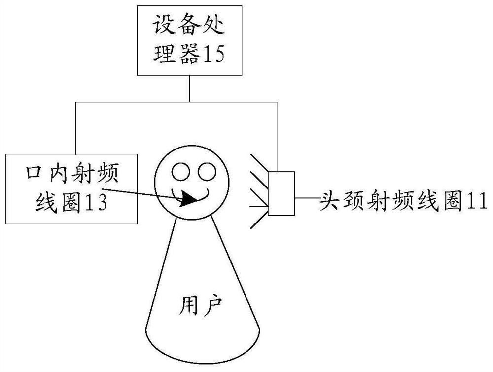
[0031] The oral craniomaxillofacial scanning equipment involved in the first embodiment of the present invention can be a low-field multifunctional oral craniomaxillofacial nuclear magnetic scanning equipment, which is small in size (because high-field nuclear magnetic equipment needs strict protection, it is difficult To achieve this, the oral craniomaxillofacial scanning device of the embodiment of the present invention adopts low field strength, small volume, low cost, low cost, and generally has a high degree of openness, low requirements for environmental protection, and can solve the problem of high field strength Existing problems), and the use of scanned MRI images to synthesize virtual CT images can solve the problem of poor hard tissue display in MRI images, and improve the accuracy of craniofacial hard tissue structure and intraoral tooth pathological analysis.

[0032] figure 1 It is a schematic diagram of an optional oral craniomaxillofacial scanning device accord...

Embodiment 2

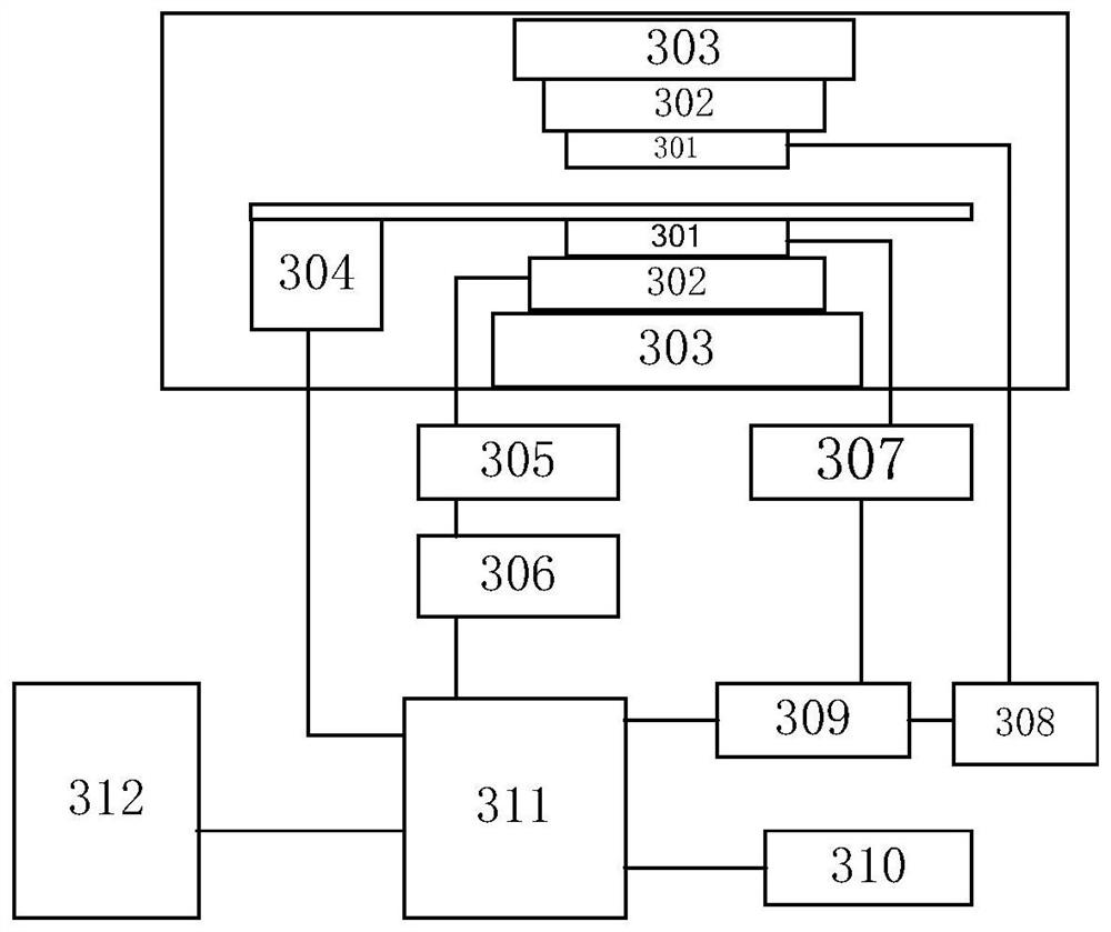
[0061] According to an embodiment of the present invention, an embodiment of an oral craniofacial scanning method is provided, and the oral craniofacial scanning method can be applied to the oral craniofacial scanning device in Embodiment 1. It should be noted that, The steps shown in the flow diagrams of the figures may be implemented in a computer system, such as a set of computer-executable instructions, and, although a logical order is shown in the flow diagrams, in some cases, may be executed differently from this The steps shown or described are performed in the order shown or described.

[0062] Figure 4 It is a flow chart of an optional oral craniomaxillofacial scanning method according to an embodiment of the present invention, such as Figure 4 As shown, the method includes the following steps:

[0063] Step S401, controlling the head and neck radio frequency coil to scan the target user's head to obtain the user's head image, wherein the user's head image is used...


PUM


Abstract

The invention discloses a scanning device, a scanning method and an electronic device for oral cavity, craniomaxillofacial area. Among them, the method includes: controlling the head and neck radio frequency coil to scan the target user's head to obtain the user's head image, wherein the user's head image is used to analyze the target user's head structure; based on the target user's head structure and head module Analyze the target user's oral craniomaxillofacial features, and obtain the oral craniomaxillofacial analysis results; control the whole dental arch current in the intraoral radio frequency coil to scan the target user's entire dentition, and obtain the first magnetic resonance image MRI; based on The first magnetic resonance image MRI synthesizes the first virtual CT image, and based on the first magnetic resonance image MRI and the first virtual CT image, analyze the tooth structure in the oral cavity of the target user, wherein the tooth structure in the oral cavity is used to determine the tooth structure in the oral cavity of the target user Dental pathology information: Based on the oral craniomaxillofacial analysis results and dental pathological information, determine whether the target user's oral craniomaxillofacial is abnormal.

Description

technical field [0001] The present invention relates to the technical field of image processing, in particular to a scanning device, a scanning method, and an electronic device for oral cavity, craniomaxillofacial area. Background technique [0002] In related technologies, in clinical imaging and 3D cephalometric research, the establishment of a 3D coordinate system and reference plane is crucial, especially with the continuous popularization of CBCT (Cone beam computed tomography, CBCT), it is easy to obtain from the outside through a computer terminal. Obtain the image information of the user's oral cavity and craniomaxillofacial area. At present, when obtaining oral craniomaxillofacial images, the commonly used imaging equipment includes curved surface tomography machine, dental film machine, CBCT machine, etc., but these equipments all use X-ray imaging, X-rays have radiation, and X-rays form There is little soft tissue information in the image, and it can only analyze...

Claims


Application Information

Patent Timeline
Patent Type & Authority Patents(China)
IPC IPC(8): A61B5/055A61B6/03A61B6/14
CPCA61B5/055A61B5/4542A61B5/4547A61B6/032A61B6/14A61B6/4417A61B6/5247
Inventor 姜喜玲付明山杨莉
Owner 赤峰学院附属医院
Why Eureka
Try Eureka