Image reconstruction method for precisely recognizing stroke intracranial lesion area

A lesion area, image reconstruction technology, applied in image data processing, 2D image generation, medical imaging and other directions, can solve the problem of limiting the application of the total variation method, reducing the overall resolution of the reconstructed image, etc., to overcome the edge over-smooth effect , the effect of improving stability and applicability, and expanding the scope of application

Pending Publication Date: 2020-09-04
FOURTH MILITARY MEDICAL UNIVERSITY
View PDF0 Cites 8 Cited by
  • Summary
  • Abstract
  • Description
  • Claims
  • Application Information

AI Technical Summary

Problems solved by technology

However, since the total variation method tends to produce a piecewise constant solution, this method also produces a "staircase effect" in smooth areas while r

Method used


Image

  • Image reconstruction method for precisely recognizing stroke intracranial lesion area
  • Image reconstruction method for precisely recognizing stroke intracranial lesion area
  • Image reconstruction method for precisely recognizing stroke intracranial lesion area

Examples


Embodiment Construction

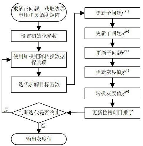
[0023] An image reconstruction method for accurately identifying intracranial lesion areas in stroke according to the present invention will be described with reference to the drawings and embodiments.

[0024] An image reconstruction method for accurately identifying intracranial lesions in stroke according to the present invention is used for EIT reconstruction of electrical conductivity distribution of hemorrhagic stroke. In order to reduce the difficulty of solving the objective function, a weighting matrix is ​​proposed to convert the data fidelity item; at the same time, in order to further remove artifacts and improve robustness, a soft threshold operator is introduced; finally, an effective iterative The algorithm completes the final solution of the inverse problem.

[0025] Such as figure 1 As shown, it is a flowchart of an image reconstruction method for accurately identifying intracranial lesion areas in stroke according to the present invention.

[0026] Such as ...


PUM


Abstract

The invention discloses an image reconstruction method for precisely recognizing a stroke intracranial lesion area and is used for reconstructing an electrical conductivity distribution of hemorrhagicstroke by electrical impedance tomography (EIT). According to the method, the EIT problem is regarded as a linear ill-posed problem and a target function is determined; and the process of the reconstruction method comprises the following steps: acquiring object field information, and solving a relative boundary measurement value vector and a sensitivity matrix; setting an initialization parameter; transforming a data fidelity item by using a weighting matrix; solving the target function in an iteration manner; judging whether iteration ends or not; and performing imaging according to an obtained gray value. The image reconstruction method effectively overcomes an edge excessive smoothness effect and inhibits stair step artifacts, improves the stability and applicability of an algorithm and the spatial resolution of a reconstructed image, and expands the application range of a total variation regularization algorithm in the medical detection field.

Description

technical field [0001] The invention belongs to the technical field of electrical impedance tomography, and in particular relates to an image reconstruction method for accurately identifying intracranial lesion areas in stroke. Background technique [0002] Currently, hemorrhagic stroke is considered a serious brain disease that exposes the brain tissue to the stimulation of blood, resulting in disability or death of the patient. In the diagnosis of hemorrhagic stroke, computed tomography (Computed Tomography, CT) and magnetic resonance imaging (Magnetic Resonance Imaging, MRI) are two commonly used imaging techniques with high spatial resolution. However, neither technique can detect the early course of hemorrhagic stroke, preventing timely treatment. In addition, the time resolution of CT and MRI is very low, the imaging time is long, the equipment is bulky, and the price is relatively expensive. [0003] Electrical impedance tomography (Electrical Impedance Tomography, ...

Claims


Application Information

Patent Timeline
IPC IPC(8): A61B5/053A61B5/00G06F17/16G06F30/23G06T11/00
CPCA61B5/0042A61B5/0536A61B5/4064A61B2576/026G06F17/16G06T11/003G06F30/23
Inventor 施艳艳饶祖广付峰王萌
Owner FOURTH MILITARY MEDICAL UNIVERSITY
Why Eureka
Try Eureka