Method for detecting novel coronavirus 2019-nCoV by multi-protein joint inspection combination

A coronavirus and combination detection technology, which is applied in the field of multi-protein joint detection combination detection of new coronavirus 2019-nCoV, can solve problems such as false positives and cross-reactions, achieve optimal design, increase sensitivity and specificity, and avoid false negatives and false positives. positive effect

Pending Publication Date: 2020-09-04
无锡市孚维尔生物医疗科技有限公司
View PDF10 Cites 11 Cited by
  • Summary
  • Abstract
  • Description
  • Claims
  • Application Information

AI Technical Summary

Problems solved by technology

The disadvantage of this method is that the lateral flow chromatography method uses one or at most two proteins for detection, and there is no cleaning process. Since the blood of individual patients contains rheumatoid factors, heterophilic antibodies, autoantibodies, and drugs and tumor cells, etc., It is easy to cause cross-reaction of the test and false positive results

Method used


Image

  • Method for detecting novel coronavirus 2019-nCoV by multi-protein joint inspection combination
  • Method for detecting novel coronavirus 2019-nCoV by multi-protein joint inspection combination
  • Method for detecting novel coronavirus 2019-nCoV by multi-protein joint inspection combination

Examples


Embodiment 1

[0069] Preparation of test strips:

[0070] 1. Reagent preparation:

[0071] 1.1 Protein dilution: Weigh 55.8g of disodium hydrogen phosphate, 5.5g of sodium dihydrogen phosphate, and 9g of sodium chloride; add 999mL of pure water to dissolve, draw 1mL of proClin300 into the solution, stir and mix, and dilute to 1000mL. Store at room temperature for later use.

[0072] 1.2 Preparation of blocking solution: Weigh 55.8 g of disodium hydrogen phosphate, 5.5 g of sodium dihydrogen phosphate, 9 g of sodium chloride, and 10 g of bovine serum albumin, add 900 mL of pure water to dissolve, add NaOH solution to adjust the pH to 7.4. Draw 1mL proClin300 into the solution, stir and mix, and dilute to 1000mL. Store at 2-8°C for later use.

[0073] 2. Preparation steps:

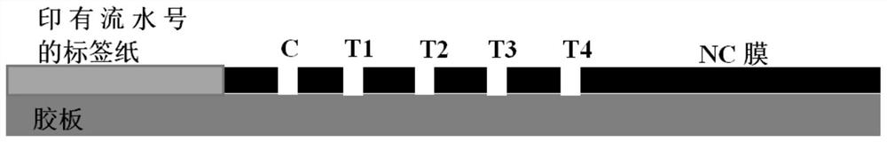
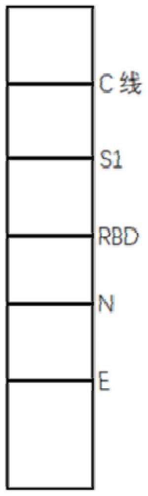
[0074] 2.1 The length and width of the nitrocellulose membrane were cut to 31cm×5cm, and four kinds of high-purity specific recombinant S1 protein, recombinant RBD protein, recombinant N protein, and recombinant E pro...

Embodiment 2

[0090] Preparation of test strips:

[0091] 1. Reagent preparation:

[0092] 1.1 Protein dilution: Weigh 55.8g of disodium hydrogen phosphate, 5.5g of sodium dihydrogen phosphate, and 9g of sodium chloride; add 999mL of pure water to dissolve, draw 1mL of proClin300 into the solution, stir and mix, and dilute to 1000mL. Store at room temperature for later use.

[0093] 1.2 Preparation of blocking solution: Weigh 55.8 g of disodium hydrogen phosphate, 5.5 g of sodium dihydrogen phosphate, and 9 g of sodium chloride; weigh 10 g of bovine serum albumin, add 900 mL of pure water to dissolve, add NaOH solution to adjust the pH to 7.4. Draw 1mL proClin300 into the solution, stir and mix, and dilute to 1000mL. Store at 2-8°C for later use.

[0094] 2. Preparation steps:

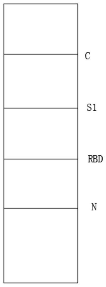
[0095] 2.1 The length and width of the nitrocellulose membrane are cut to 31cm×5cm, and three kinds of high-purity specific recombinant S1 protein, recombinant RBD protein, and recombinant N protein are respecti...

Embodiment 3

[0111] Preparation of test strips:

[0112] 1. Reagent preparation:

[0113] 1.1 Protein dilution: Weigh 55.8g of disodium hydrogen phosphate, 5.5g of sodium dihydrogen phosphate, and 9g of sodium chloride; add 999mL of pure water to dissolve, draw 1mL of proClin300 into the solution, stir and mix, and dilute to 1000mL. Store at room temperature for later use.

[0114] 1.2 Preparation of blocking solution: Weigh 55.8 g of disodium hydrogen phosphate, 5.5 g of sodium dihydrogen phosphate, and 9 g of sodium chloride; weigh 10 g of bovine serum albumin, add 900 mL of pure water to dissolve, add NaOH solution to adjust the pH to 7.4. Draw 1mL proClin300 into the solution, stir and mix, and dilute to 1000mL. Store at 2-8°C for later use.

[0115] 2. Preparation steps:

[0116] 2.1 The length and width of the nitrocellulose membrane are cut to 31cm×5cm, and three kinds of high-purity specific recombinant S1 protein, recombinant RBD protein, and recombinant E protein are respecti...


PUM

PropertyMeasurementUnit
widthaaaaaaaaaa
lengthaaaaaaaaaa
lengthaaaaaaaaaa

Abstract

The invention relates to a method for detecting novel coronavirus 2019-nCoV through multi-protein joint inspection combination, which belongs to the technical field of novel coronavirus 2019-nCoV detection. The invention provides the method for detecting the novel coronavirus 2019-nCoV through multi-protein joint inspection combination. A detection test strip for detecting the novel coronavirus 2019-nCoV antibody is prepared by directly scribing a high-purity and specific novel coronavirus antigen on a nitrocellulose membrane, and IgG, IgM and / or IgA antibodies in a sample can be simultaneously detected by utilizing multiple antigens. Through joint inspection of three or four specific antigens, protein sequences of the antigens are optimally designed, so that the sensitivity and specificity are effectively improved, and the problems of false negative and false positive are effectively avoided.

Description

technical field [0001] The invention relates to a method for detecting novel coronavirus 2019-nCoV by a multi-protein joint detection combination, belonging to the technical field of novel coronavirus 2019-nCoV detection. Background technique [0002] Coronaviruses are a large family of viruses known to cause colds as well as more serious illnesses such as Middle East Respiratory Syndrome (MERS) and Severe Acute Respiratory Syndrome (SARS). Novel coronavirus is a new strain of coronavirus that has never been found in humans before, and the World Health Organization named the disease caused by this coronavirus as coronavirus disease 2019 (COVID-19), the deadly virus severe acute respiratory syndrome Coronavirus type 2 (SARS-CoV-2). [0003] The coronavirus genome encodes spike protein (spike protein), envelope protein (Envelopeprotein, E protein), membrane protein (Membrane protein) and nucleocapsid protein (Nucleocapsid, N protein) in sequence. Spike protein is the most im...

Claims


Application Information

Patent Timeline
Patent Type & Authority Applications(China)
IPC IPC(8): G01N33/68G01N33/58G01N33/569G01N33/558
CPCG01N33/6854G01N33/581G01N33/56983G01N33/558G01N2333/165G01N2469/20
Inventor 吴玉章吴长龙
Owner 无锡市孚维尔生物医疗科技有限公司
Why Eureka
Try Eureka