Supercharge Your Innovation With Domain-Expert AI Agents!

Cancer hyperspectral image segmentation method and system based on double-branch attention deep learning

A hyperspectral image and deep learning technology, applied in the field of cancer hyperspectral image segmentation methods and systems, can solve the problems of poor robustness, inability to extract spatial attention information and channel attention information of hyperspectral images, and low efficiency. Robust effect

Active Publication Date: 2020-09-15
EAST CHINA NORMAL UNIV
View PDF5 Cites 18 Cited by
  • Summary
  • Abstract
  • Description
  • Claims
  • Application Information

AI Technical Summary

Problems solved by technology

[0006] Although the existing hyperspectral image processing methods can achieve hyperspectral image segmentation, there are still three deficiencies in the processing flow: First, the existing methods cannot extract hyperspectral images on multiple scales when extracting hyperspectral features. Spatial attention information and channel attention information of
The commonly used attention mechanism often only extracts features on a single scale, but there is no corresponding strategy for hyperspectral images with different scale features
Second, the existing network structure cannot extract image features on a larger spatial scale.
However, cancer hyperspectral images have large-scale morphological features due to factors such as tissue or cell structure and shape, so the method of using image blocks as network input is not applicable.
Third, in the case of using traditional machine learning methods, the processing flow is often not an end-to-end model, resulting in loss of accuracy of results and complicated processing steps
SVM is a commonly used machine learning method. However, when dealing with hyperspectral images, it is often necessary to first perform dimensionality reduction operations on the data through the PCA method. However, since the model is not an end-to-end structure, the dimensionality reduction strategy of PCA and the method of segmenting hyperspectral images The goals are different, so some important information that is helpful for segmentation may be lost during the dimensionality reduction process, and the entire model cannot be optimized
These three deficiencies will lead to less robustness and lower efficiency in practical hyperspectral image segmentation problems

Method used


Image

  • Cancer hyperspectral image segmentation method and system based on double-branch attention deep learning
  • Cancer hyperspectral image segmentation method and system based on double-branch attention deep learning
  • Cancer hyperspectral image segmentation method and system based on double-branch attention deep learning

Examples


Embodiment

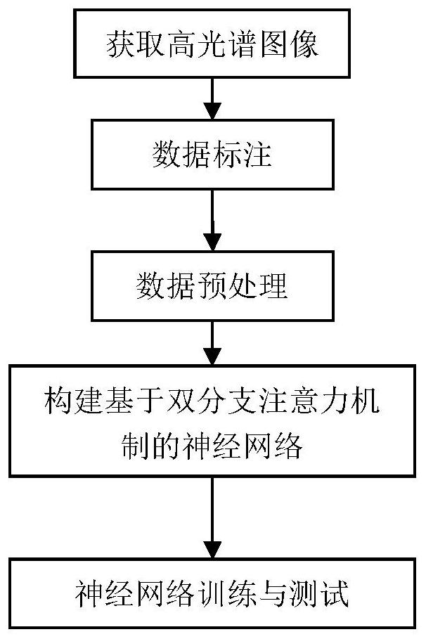
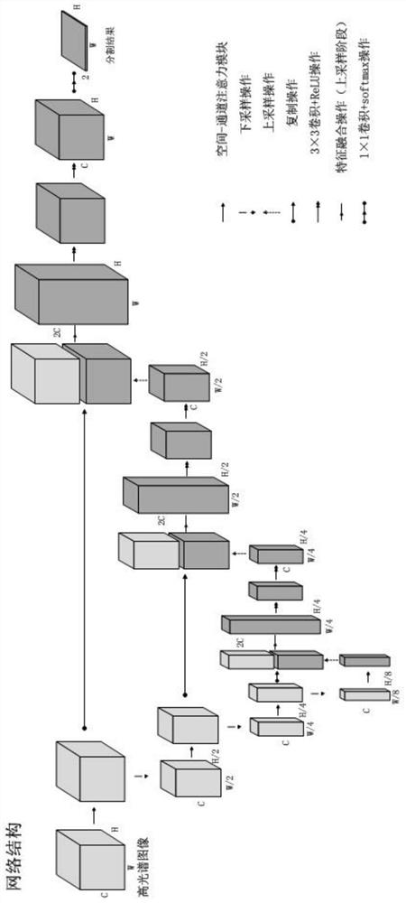
[0102] This embodiment takes cholangiocarcinoma as an example to illustrate the conditions, processes, steps, principles and results of the present invention. refer to figure 1 and figure 2 , figure 1 is a flowchart of a cancer hyperspectral image segmentation method using dual-branch attention-based deep learning, figure 2 is the network structure of the segmentation method.

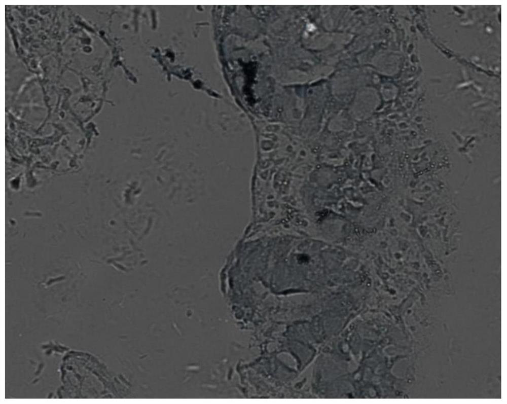
[0103] Acquire hyperspectral images and annotate and preprocess them. Hyperspectral images can be collected by hyperspectral microscopes, but due to the acquisition equipment, noise will inevitably be introduced, and the contrast of the image in a specific spectral band needs to be adjusted. Therefore, the median filter and image normalization methods are respectively introduced in the preprocessing stage to process the acquired hyperspectral images. image 3 is the preprocessed single-band image of cholangiocarcinoma, Figure 4 is the corresponding labeled binary image.

[0104] Due to the lar...


PUM


Abstract

The invention discloses a cancer hyperspectral image segmentation method based on double-branch attention deep learning. The method comprises the steps: firstly carrying out the preprocessing of a cancer hyperspectral image, and dividing a data set into a training set and a test set; then constructing a double-branch deep convolutional neural network based on an attention mechanism, and training the network through the divided training set; and then the trained neural network is used to test the test set, and finally the purpose of segmenting the cancer hyperspectral image is achieved. According to the method, a double-branch structure is constructed according to the characteristics of the cancer hyperspectral image, spatial information and spectral information of the hyperspectral image are extracted respectively and fused, and finally a prediction result is obtained. The invention further discloses a cancer hyperspectral image segmentation system based on double-branch attention deeplearning.

Description

technical field [0001] The present invention relates to the technical fields of hyperspectral image processing and medical image processing, in particular to a cancer hyperspectral image segmentation method and system based on dual-branch attention deep learning. Background technique [0002] Carcinoma refers to malignant tumors originating from epithelial tissue, and is the most common type of malignant tumors. Most types of cancer lack typical clinical manifestations and diagnostic methods in the early stage. When patients have typical symptoms, they have basically entered the middle and late stages and lost the opportunity for radical surgery. There are many diagnostic methods for cancer, mainly including clinical diagnosis, physical and chemical diagnosis, surgical diagnosis, cytopathological diagnosis, and histopathological diagnosis. Histopathological diagnosis can understand the benign and malignant nature of the tumor, judge the prognosis of cancer, provide direct h...

Claims


Application Information

Patent Timeline
IPC IPC(8): G06T7/11G06K9/62G06N3/04G06N3/08
CPCG06T7/11G06N3/08G06T2207/10056G06T2207/20032G06T2207/20081G06T2207/20084G06T2207/30096G06N3/047G06N3/045G06F18/241G06F18/2415
Inventor 邱崧惠思远李庆利周梅胡孟晗徐伟
Owner EAST CHINA NORMAL UNIV
Features
  • R&D
  • Intellectual Property
  • Life Sciences
  • Materials
  • Tech Scout
  • Unparalleled Data Quality
  • Higher Quality Content
  • 60% Fewer Hallucinations
Learn More