Tumor interstitial ratio judgment method and system based on image processing algorithm

A tumor stroma and image processing technology, applied in the field of medical image processing, can solve problems such as large uncertainties

Pending Publication Date: 2020-10-23
复影(上海)医疗科技有限公司
View PDF1 Cites 3 Cited by
  • Summary
  • Abstract
  • Description
  • Claims
  • Application Information

AI Technical Summary

Problems solved by technology

[0005] At present, the calculation of the tumor cell-to-stroma ratio in major hospitals is still based on judging tumor images with the naked eye of doctors, and predicting a tumor-to-stro...

Method used


Image

  • Tumor interstitial ratio judgment method and system based on image processing algorithm
  • Tumor interstitial ratio judgment method and system based on image processing algorithm
  • Tumor interstitial ratio judgment method and system based on image processing algorithm

Examples


Embodiment 1

[0066] A method for judging the tumor-stroma ratio based on an image processing algorithm according to the present invention includes:

[0067] Step M1: Read HE immunohistochemical images of tumor pathological sections;

[0068] Step M2: Select an image whose average gray value and blur degree of the image are within a preset range;

[0069] Step M3: Preprocessing the selected image based on the image preprocessing algorithm to obtain the preprocessed image;

[0070] Step M4: Segment the preprocessed image;

[0071] Step M5: Obtain and mark the segmentation results, and calculate the mass ratio between tumors.

[0072] Specifically, the step M3 includes:

[0073] Step M3.1: Perform grayscale processing on the HE immunohistochemical image, and normalize the image data;

[0074] Step M3.2: After the image is normalized, the image is denoised for the first time by median filtering, which can better eliminate noise and make each area of ​​the image smooth;

[0075] Step M3.3:...

Embodiment 2

[0105] Embodiment 2 is a modification of embodiment 1

[0106] The purpose of the present invention is to provide a reliable solution for calculating the ratio of tumor cells to the interstitium, by dividing the interstitial part and the substantial part of the pathological HE immunohistochemical image of tumor cells, and calculating the ratio of each to the total area of ​​the image to obtain these data. This solution is more efficient and accurate than the traditional method used by doctors to calculate the ratio of tumor to stroma. At the same time, the system comes with a set of interactive GUI interface, which is user-friendly.

[0107] The present invention provides two segmentation schemes, both of which require four preprocessing steps on the image. First of all, we need to grayscale the image for two reasons: 1. The tumor-stroma ratio image is an RGB image consisting of three color channels (in Matlab, it is expressed as a three-channel matrix), which is inconvenient...

Embodiment 3

[0144] Embodiment 3 is a modification of embodiment 1 and / or embodiment 2

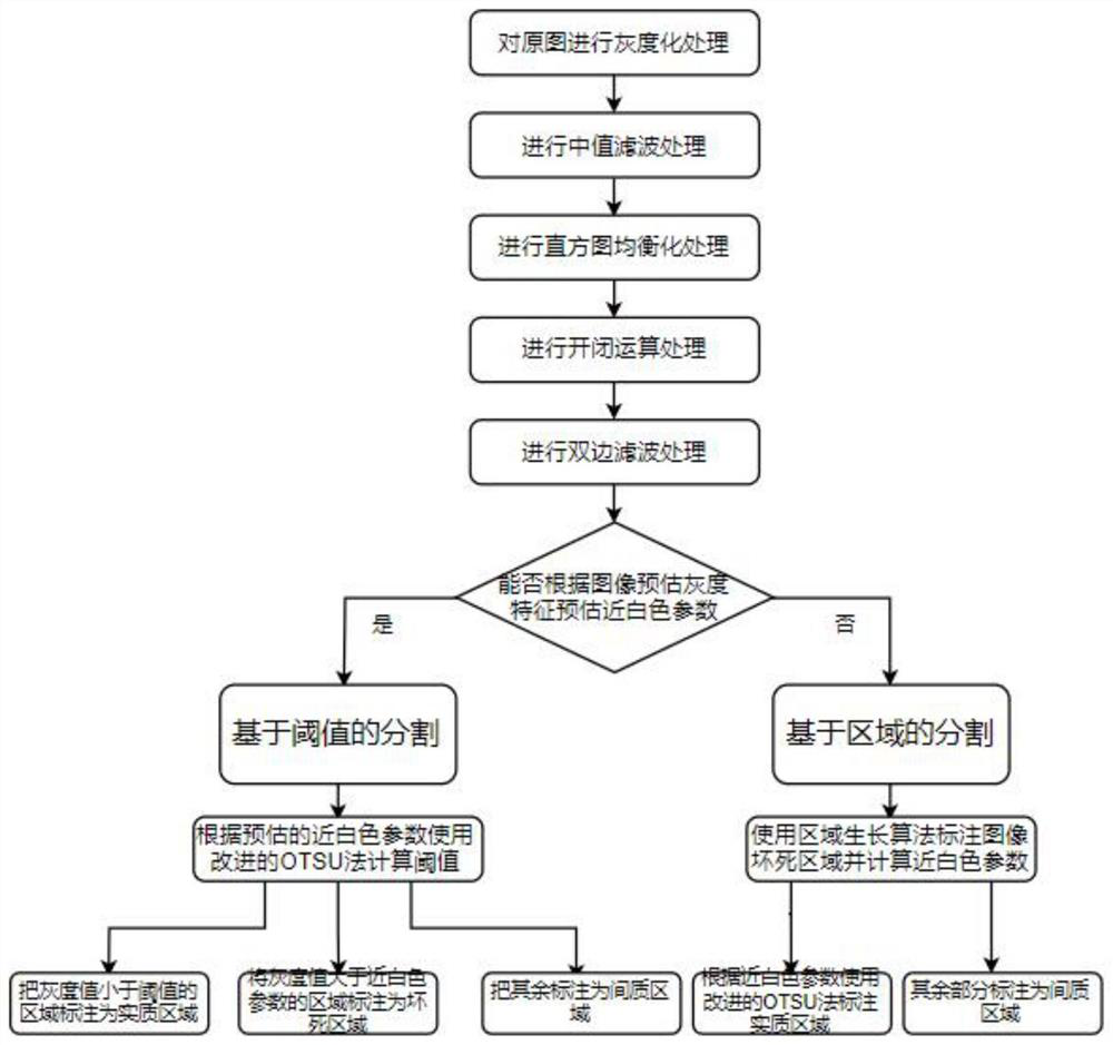
[0145] figure 1 It is a flow chart of the method of the present invention.

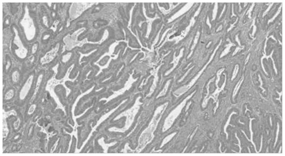
[0146] figure 2 It is the original image of the tumor image, the color distribution of the image is uneven, and it is greatly affected by noise. Therefore, it is necessary to properly preprocess the image.

[0147] Step 1: Perform grayscale processing on the original image. Grayscale processing is to scale the grayscale values ​​of all pixels in the image between [0,1]. It is a normalized process, which is convenient. The calculation of the data, and the subsequent preprocessing steps, whether it is median filtering or histogram equalization, require the gray value of the image to be controlled between [0,1]. This step is the premise of the algorithm.

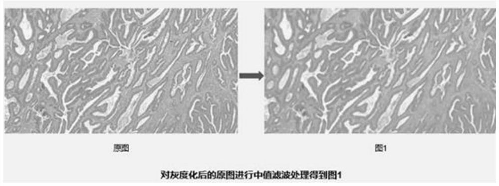
[0148] Step two, such as image 3 As shown, median filtering is performed on the grayscaled image for the first noise reduction. Median filtering (Median filtering) is an ...


PUM


Abstract

The invention provides a tumor interstitial ratio judgment method and system based on an image processing algorithm. The tumor interstitial ratio judgment method comprises the steps OF: M1, reading tumor pathological section HE immunohistochemical images; M2, selecting an image of which the average gray value and of which the blurring degree are within a preset range; M3, preprocessing the selected image based on an image preprocessing algorithm to obtain a preprocessed image; M4, segmenting the preprocessed image is; and M5, obtaining a segmentation result, marking the segmentation result, and calculating the mass ratio between tumors. According to the method, the accuracy of data is greatly improved, and a set of tumor interstitial ratio calculation mode with higher efficiency and higherprecision is provided.

Description

technical field [0001] The present invention relates to the field of medical image processing, in particular, to a method and system for judging tumor-stroma ratio based on image processing algorithms, and more specifically, to a method and system for fast pathological slice image-stroma ratio calculation, and to Calculation of data such as the stroma ratio of tumor pathological HE immunohistochemical images; in particular, an algorithm for segmenting and labeling the stroma and parenchyma of tumor pathological HE immunohistochemical images. Background technique [0002] In 2018, there were 18.1 million new cancer patients worldwide, and 9.6 million cancer deaths. my country is a country with a large population and a high incidence of cancer. In 2018, the number of new cases in my country was 3.804 million, accounting for more than 20% of the number of new cancer cases in the world. , equivalent to an average of more than 10,000 people are diagnosed with cancer every day, an...

Claims


Application Information

Patent Timeline
IPC IPC(8): G06T7/00G06T5/00G06T5/20G06T5/40G06T7/11G06T7/136G06T7/90
CPCG06T7/0012G06T5/002G06T5/20G06T5/40G06T7/11G06T7/136G06T7/90G06T2207/20032G06T2207/20028G06T2207/30096G06T2207/30024
Inventor 耿道颖于泽宽陈子昂李郁欣张军尹波吴昊耿岩胡斌陆逸平李海庆周书怡曹鑫朴思蓉
Owner 复影(上海)医疗科技有限公司
Why Eureka
Try Eureka