Preparation method of chitosan derivative nanoparticles for delivering siRNA

A technology of chitosan derivatives and nanoparticles, applied in the field of gene biology, to achieve the effect of simple method and good transfection effect

Active Publication Date: 2020-10-30
HEFEI UNIV OF TECH
View PDF1 Cites 2 Cited by
  • Summary
  • Abstract
  • Description
  • Claims
  • Application Information

AI Technical Summary

Problems solved by technology

At present, there are no nanoparticles formed by encapsulating siRNA with carboxymethyl chitosan and chitosan hydrochloride, which can prom

Method used


Image

  • Preparation method of chitosan derivative nanoparticles for delivering siRNA
  • Preparation method of chitosan derivative nanoparticles for delivering siRNA
  • Preparation method of chitosan derivative nanoparticles for delivering siRNA

Examples


Embodiment 1

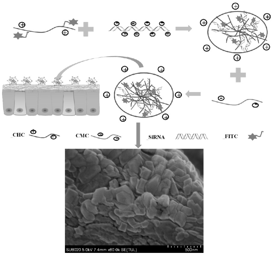
[0025] The specific steps of preparing FITC-labeled chitosan hydrochloride powder are as follows:

[0026] 10 mg of chitosan hydrochloride was completely dissolved in 10 mg of deionized water to obtain a chitosan hydrochloride solution with a concentration of 1 mg / mL. Add 10 mL of 5.8 mg / L fluorescein isothiocyanate (FITC) solution (5.8 mg / L, anhydrous methanol) to the chitosan hydrochloride solution with a concentration of 1 mg / mL, and stir under a magnetic force (500 rpm). ) For 4 h. Then, 1 M hydrogen hydroxide (NaOH) was added to adjust the pH to 10, and FITC-labeled chitosan hydrochloride was precipitated. The free FITC was washed with ultrapure water and centrifuged until no fluorescence was detected in the supernatant. Finally, the FITC-labeled chitosan hydrochloride powder was obtained by freeze-drying.

[0027] Prepare siRNA-CDNPs according to the siRNA, different volumes of CHC and different volumes of CMC (600μL, 500μL, 400μL, 300μL) in Table 1. The preparation steps ...

Embodiment 2

[0033] Application of siRNA-CDNPs in simulating colon cancer cell environment siRNA controlled release:

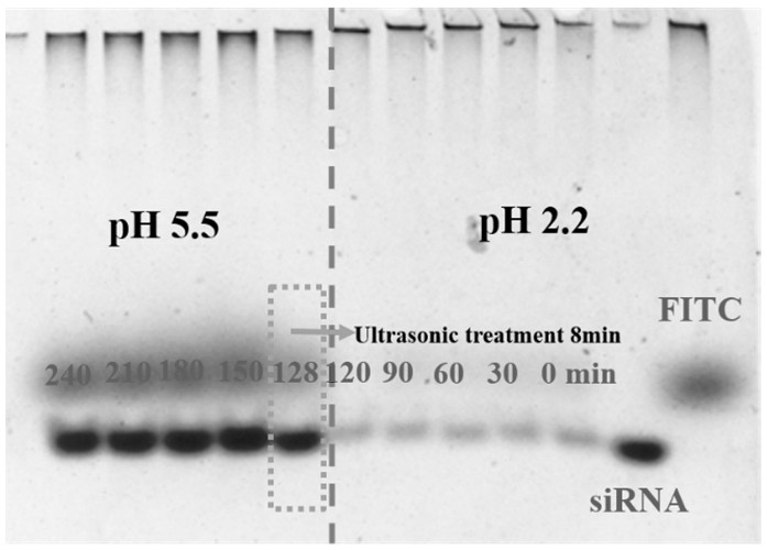
[0034] Take the siRNA-CDNPs prepared in Example 1 and incubate them in a gastric environment (pH = 2.2, 120 rpm) at 37°C for different times, up to 120 min. Then mix the above acidic solution with NaOH (1 M) and adjust the pH to a weak acid (pH = 5.5). Then sonicate for 8 min, incubate at 37°C, shake well (120 rpm) for 112 min. Obtain samples at predetermined times (0, 30, 60, 90, 120, 128, 150, 180, 210, and 240 min). The samples were analyzed by 5% (w / v) polyacrylamide gel, 120 mV electrophoresis in 1×TBE buffer, the gel was stained with ethidium bromide, and the gel imager FLA 7000 was used to observe the results. figure 2 Shown.

[0035] By analyzing the siRNA content at pH 2.2 and pH 5.5, polyacrylamide gel electrophoresis was used to detect the release of siRNA in siRNA-CDNPs. figure 2 The red arrow and white arrow in indicate unencapsulated siRNA and FITC. The rele...

Embodiment 3

[0037] Application of siRNA-CDNPs that are effectively internalized by HT-29 cells:

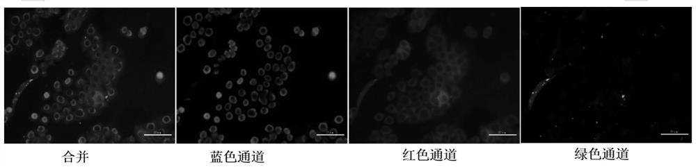
[0038] Take the siRNA-CDNPs prepared in Example 1 and incubate colon cancer cells (HT-29) to analyze the transfection effect of siRNA. The specific steps are as follows: 100μL siRNA-CDNPs (containing 0.55 nmol siRNA) are added to HT-29 cells (1×10 5 / Well) incubate for 16 h. Then the added siRNA-CDNPs were gently absorbed, and the cells were washed three times with PBS. Cells were fixed with 4% paraformaldehyde PBS for 15 min, washed twice with PBS, and nuclear stained with DAPI (4′,6-diamino-2-phenylindole) for 10 min. The cells were washed again with PBS, rinsed with ultrapure water, and placed on a microscope slide. Take a fluorescence image with a fluorescence microscope, the results are as follows image 3 Shown.

[0039] The siRNA-CDNPs prepared in Example 1 were added to colon cancer cells (HT-29), and the transfection effect of siRNA was analyzed. The results are as follows image 3 Sho...


PUM


Abstract

The invention relates to a preparation method of chitosan derivative nanoparticles for delivering siRNA, and belongs to the technical field of gene biology. A positively charged chitosan hydrochloridesolution can be mixed with an siRNA solution capable of inhibiting protein expression related to proliferation and migration of colon cancer cells, then a carboxymethyl chitosan solution with negative electricity is added, and self-assembly is performed to form chitosan derivative nanoparticles (siRNA-CDNPs) for delivering siRNA through electrostatic interaction between molecules. The chitosan derivative nanoparticles for delivering the siRNA can effectively protect the siRNA under the condition that the pH value is 1.2, and under the condition that the pH value of a colon cancer cell environment is 5.5, controllable release of the siRNA is realized through response to external stimulation (ultrasound); the siRNA-CDNPs can effectively inhibit the expression of beta-catenin protein in colon cancer cells, and is expected to inhibit the proliferation and migration of cancer cells. The preparation method is easy to operate, green, environment-friendly and low in cost.

Description

Technical field [0001] The invention belongs to the technical field of gene biology, and specifically relates to the preparation and application of a pH-sensitive siRNA / chitosan derivative nano system. Background technique [0002] Gene-based therapies have great potential in regulating the silencing of a large number of target mRNA molecules, and ultimately can reduce the level of target proteins. However, the application of this gene silencing technology still needs to overcome challenges, including the rapid degradation of siRNA under physiological conditions and the difficulty of passing through the negatively charged cytoplasmic membrane. The key challenge to realize the therapeutic efficacy of siRNA is to develop and design a new type of degradable vector that has a safe and sufficient gene delivery efficiency. Currently, the 5-year survival rate of patients undergoing chemotherapy or resection of colon cancer liver metastases is less than 50%. It is generally believed th...

Claims


Application Information

Patent Timeline
IPC IPC(8): A61K48/00A61K31/713A61K9/51A61K47/69A61P35/00A61P35/04
CPCA61K31/713A61K9/5161A61K47/6939A61P35/00A61P35/04
Inventor 郑磊颜玲刘长虹高胜杰刘帅
Owner HEFEI UNIV OF TECH
Why Eureka
Try Eureka