
CT image auxiliary diagnosis system based on cascade connection

A CT imaging and auxiliary diagnosis technology, applied in the field of image processing, can solve the problems of fatigue, large workload of CT image screening, false detection and missed detection, and achieve the effect of reducing workload and improving the quality of lesion detection.

Pending Publication Date: 2020-11-10
CHONGQING UNIV +1
View PDF7 Cites 2 Cited by
  • Summary
  • Abstract
  • Description
  • Claims
  • Application Information

AI Technical Summary

Problems solved by technology

[0004] In view of this, in order to solve the above key problems, the object of the present invention is to provide a cascade-based CT image-aided diagnosis system, which helps to solve the problem of manual CT image screening with heavy workload, fatigue, false detection and missed detection. inspection problem

Method used


Image

  • CT image auxiliary diagnosis system based on cascade connection
  • CT image auxiliary diagnosis system based on cascade connection
  • CT image auxiliary diagnosis system based on cascade connection

Examples


Embodiment Construction

[0029] The present invention will be further described below in conjunction with the accompanying drawings and embodiments.

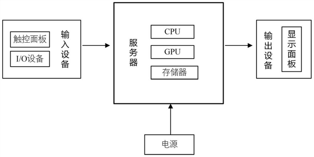
[0030] This embodiment is based on a cascaded CT image-aided diagnosis system, which includes a server end, the server end includes a server and an input device and an output device connected to the server, and an image preprocessing module and a cascade detection model are stored in the server .

[0031] The input device includes an I / O device, and the server can obtain CT images from the input device, and send the processing result of the image to the output device.

[0032] The image preprocessing module is used to convert the original CT picture into a format acceptable to the neural network. The image preprocessing module implements the following steps when being called and executed by the server:

[0033] 1) Obtain and read the initial medical CT image from the input device, and convert the original CT image saved in DICOM format into a 16-bit p...


PUM


Abstract

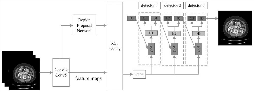
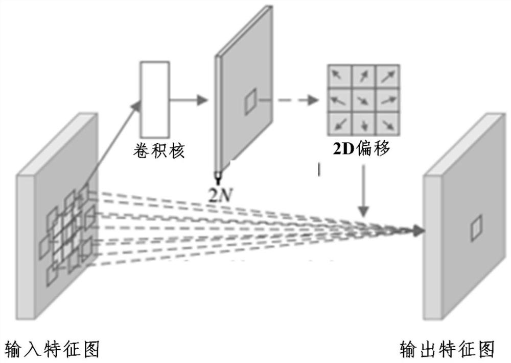
The CT image auxiliary diagnosis system based on cascade connection comprises a server side, the server side comprises a server, input equipment and output equipment, the input equipment and the output equipment are connected with the server, and an image preprocessing module and a cascade connection detection model are stored in the server; the cascade detection model comprises a convolutional neural network, a region suggestion network, an ROI Pooling layer and a plurality of cascade detectors. According to the invention, medical imaging and computer image processing are combined to completeautomatic detection of lesions. Aiming at the problem that CT focus features are complex and difficult to extract, a deformable convolution kernel is introduced into the cascade detection model to replace a standard convolution kernel, that is, 2D offset is increased in the standard convolution kernel, so that the focus features can be better extracted. Experimental results show that the lesion detection quality can be effectively improved, doctors can be helped to finish case classification and lesion identification more quickly and accurately, and the workload of the doctors is reduced.

Description

technical field [0001] The invention relates to the technical field of image processing, in particular to a CT image processing device. Background technique [0002] With the aging of the population, industrialization, the intensification of urbanization, and changes in lifestyles, the incidence of cancer is gradually increasing. Nowadays, cancer has become one of the main obstacles affecting our quality of life and health, especially lung cancer and lymphoma. This is because most patients with lung cancer and lymphoma have no obvious special symptoms in the early stage, so they will not be discovered until the late stage of cancer when they go to the hospital for examination. At this time, the best period for treatment and rehabilitation has been missed. With the development of medical imaging, doctors can use CT images to make timely diagnosis of cancer at an early stage, thereby solving the problem of high cancer mortality. With the emergence of a large number of CT ima...

Claims


Application Information

Patent Timeline
Patent Type & Authority Applications(China)
IPC IPC(8): G16H50/20G16H30/20G06T7/00
CPCG16H50/20G16H30/20G06T7/0012G06T2207/10081G06T2207/20081G06T2207/20084G06T2207/30096
Inventor 刘然赵洋易琳刘亚琼陈希崔珊珊王斐斐陈丹
Owner CHONGQING UNIV
Learn More