Skin cancer lesion segmentation method based on deep learning

A deep learning and skin cancer technology, applied in neural learning methods, image analysis, image data processing, etc., can solve the problems of further improvement of segmentation effect, blurred skin of lesions caused by background interference, and large changes in scale of skin of lesions, etc. Output segmentation results, high-efficiency semantic segmentation, and the effect of suppressing interference

Active Publication Date: 2020-11-17
NANHUA UNIV
View PDF2 Cites 7 Cited by
  • Summary
  • Abstract
  • Description
  • Claims
  • Application Information

AI Technical Summary

Problems solved by technology

Due to the complexity of the dermoscopic image itself, although the convolutional neural network can well complete the semantic segmentation task of natural images, its application in the field of dermoscopic image segmentation is immature, and the segmentation effect has room for further improvement.
Due to some challenges in the dermoscopic image: large scale changes of the lesion skin, more background noise in the image, and blurred edges of the lesion skin

Method used


Image

  • Skin cancer lesion segmentation method based on deep learning
  • Skin cancer lesion segmentation method based on deep learning
  • Skin cancer lesion segmentation method based on deep learning

Examples


Embodiment Construction

[0041] The implementation of the present invention will be illustrated by specific specific examples below, and those skilled in the art can easily understand other advantages and effects of the present invention from the content disclosed in this specification. The structures or working principles not described in detail in the present invention belong to the prior art and common knowledge in the field, and should be known to those skilled in the art.

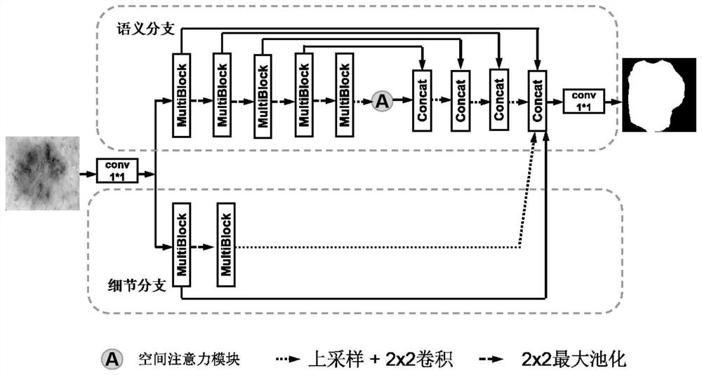
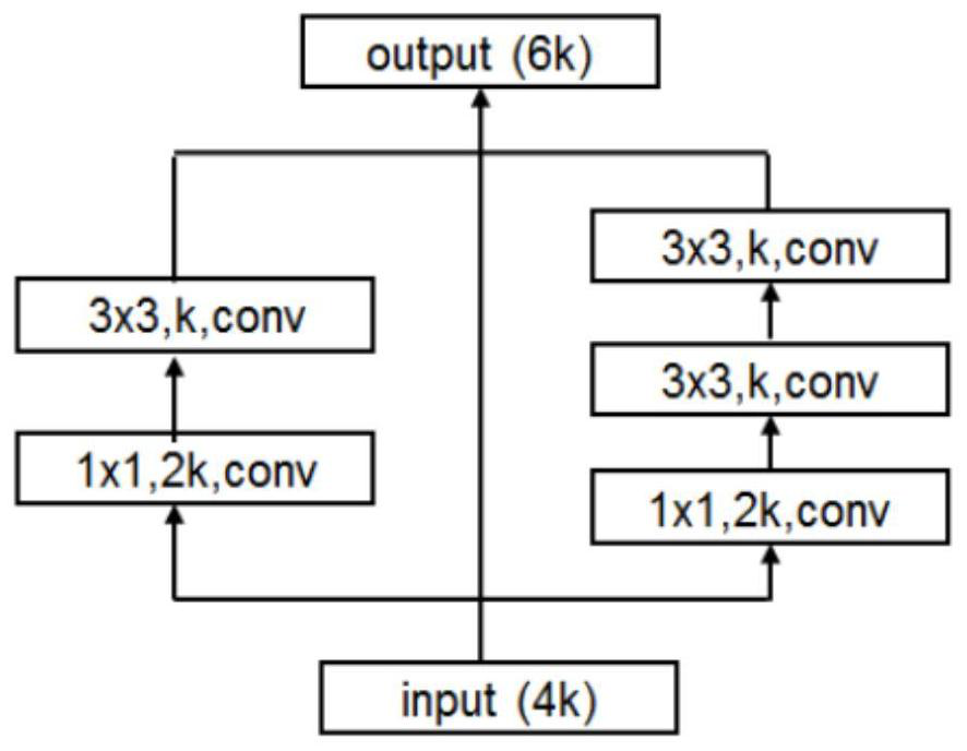
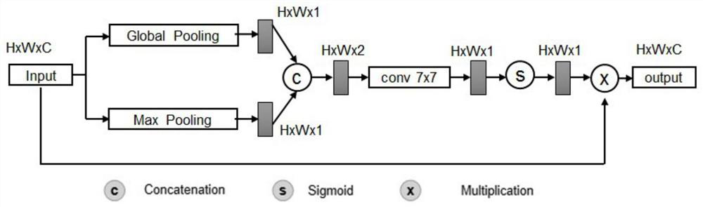
[0042] The present invention is realized under the Keras deep learning framework, and the computer configuration adopts: Intel Core i5 6600K processor, 16G memory, NVIDIA V100 graphics card, Linux operating system. The present invention provides a skin cancer lesion segmentation method based on deep learning, which specifically includes the following steps:

[0043] Step 1, obtain training dermoscopic image samples:

[0044]The dermoscopic images come from the International Skin Open Challenge dataset (ISIC 2018), which conta...


PUM


Abstract

The invention discloses a skin cancer lesion segmentation method based on deep learning. The method comprises the steps of 1, obtaining a training dermatoscope image sample; 2, normalizing data; 3, designing an edge perception neural network model; 4, training the edge perception neural network model; 5, performing segmentation. Shallow detail information and deep semantic information are fused, edge details of images can be well detected. A Multi-Block module is used for expanding the receptive field of the model so as to enhance the sensitivity to targets of different scales, and meanwhile,the interference of background information is suppressed in combination with a spatial attention mechanism.

Description

technical field [0001] The invention relates to the technical field of computer-aided diagnosis, in particular to a method for segmenting skin cancer lesions based on deep learning. Background technique [0002] Skin cancer and various pigmented skin diseases are seriously threatening human health. At present, the medical field mainly realizes the diagnosis of pigmented skin diseases through the observation and analysis of the lesion characteristics in the dermoscopic images by doctors. Dermoscopy image is a medical image obtained by non-invasive microscopic imaging technology, which can clearly show the lesion characteristics of skin diseases. However, due to the small differences in lesions in different cases, it is very difficult for doctors to analyze and judge the type of lesions by naked eye observation. In order to achieve effective treatment, the demand for computer-aided diagnosis systems for dermoscopic images is increasing. Computer-aided diagnosis can relieve t...

Claims


Application Information

Patent Timeline
Patent Type & Authority Applications(China)
IPC IPC(8): G06T7/12G06T7/136G06T7/181G06T7/187G06N3/04G06N3/08
CPCG06T7/12G06T7/136G06T7/181G06T7/187G06N3/08G06T2207/10004G06T2207/10024G06T2207/20081G06T2207/20084G06T2207/30088G06N3/048G06N3/045Y02T10/40
Inventor 屈爱平程志明梁豪钟海勤黄家辉
Owner NANHUA UNIV
Why Eureka
Try Eureka