Method for intracellular delivery of protein

A protein and protein solution technology, applied in the field of bioengineering, can solve problems such as easy to be removed, protein cannot function, CPPs short half-life, etc., to achieve the effect of avoiding chemical bond modification, excellent biocompatibility, and simple operation steps

Inactive Publication Date: 2020-12-15
CHINA UNIV OF PETROLEUM (EAST CHINA)
View PDF7 Cites 1 Cited by
  • Summary
  • Abstract
  • Description
  • Claims
  • Application Information

AI Technical Summary

Problems solved by technology

However, the half-life of CPPs is short and is easy to be cleared (D.Sarko, B.Beijer, R.Garcia Boy, et al.The Pharmacokinetics of Cell-Penetrating Peptides[J].Molecular Pharmaceuticals,2010,7(6):2224 -2231.)
At the same time, a considerable proportion of CPPs will be wrapped in endosomes after internalization, which directly leads to the inability of the proteins carried by them to function, and is bound in endosomes for a long time. The acidic environment after endosomes combine with lysosomes and various The enzyme will further degrade the protein (J.C.LeCher, S.J.Nowak, J.L.McMurry. Breaking in and busting out: cell-penetrating peptides and the endosomal escape problem[J].Biomolecular concepts,2017,8(3-4):131-141. )

Method used


Image

  • Method for intracellular delivery of protein
  • Method for intracellular delivery of protein
  • Method for intracellular delivery of protein

Examples


Embodiment 1

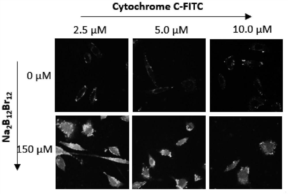
[0023] (1) Take boron halide clusters (Na 2 B 12 Br 12 ) and labeled cytochrome C, the solvent is 1×PBS, the total volume is 100 μL, mixed thoroughly for 10 min, and sterilized by filtration through a 0.22 μm microporous membrane;

[0024] (2) Add 300 μL DMEM to the 24-well plate where NIH-3T3 cells were cultured, and add 100 μL of the mixture in step (1), incubate for 6 hours, boron halide clusters (Na 2 B 12 Br 12 ) with a final concentration of 150 μM, and labeled cytochrome C concentrations of 2.5 μM, 5.0 μM and 10.0 μM;

[0025] (3) Aspirate the mixture in the 24-well plate in step (2), wash with 1×PBS three times, stain and fix the cells;

[0026] (4) Carry out confocal imaging of the cells using a confocal microscope to observe the distribution of labeled cytochrome C in the cells. The distribution of proteins in the cells can be seen in the fluorescent channel of 500-530nm labeled cytochrome C, from figure 2 Boron halide clusters (Na 2 B 12 Br 12 ) can effici...

Embodiment 2

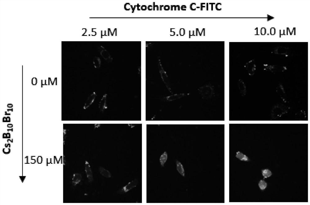
[0028] (1) boron halide clusters (Cs) with a mass ratio of about (1~5):1 2 B 10 Br 10 ) and labeled cytochrome C, the solvent is 1×PBS, the total volume is 100 μL, mixed thoroughly for 10 min, and sterilized by filtration through a 0.22 μm microporous membrane;

[0029] (2) Add 300 μL DMEM to the 24-well plate where NIH-3T3 cells were cultured, and add 100 μL of the mixture in step (1), incubate for 6 hours, boron halide clusters (Cs 2 B 10 Br 10 ) with a final concentration of 150 μM, and labeled cytochrome C concentrations of 2.5 μM, 5.0 μM and 10.0 μM;

[0030] (3) Aspirate the mixture in the 24-well plate in step (2), wash with 1×PBS three times, stain and fix the cells;

[0031] (4) Carry out confocal imaging of the cells using a confocal microscope to observe the distribution of labeled cytochrome C in the cells. The distribution of proteins in the cells can be seen in the fluorescent channel of 500-530nm labeled cytochrome C, from image 3 boron halide clusters (C...

Embodiment 3

[0033] (1) Take boron halide clusters (Na 2 B 12 Br 12 ) and labeled cytochrome C, the solvent is 1×PBS, the total volume is 100 μL, mixed thoroughly for 20 minutes, and sterilized by filtration through a 0.22 μm microporous membrane;

[0034] (2) Add 300 μL DMEM to the 24-well plate in which HeLa cells were cultured, and add 100 μL of the mixture in step (1), incubate for 6 hours, boron halide clusters (Na 2 B 12 Br 12 ) final concentration is 200 μM, and the concentration of labeled cytochrome C is 1.0 μM;

[0035] (3) Aspirate the mixture in the 24-well plate in step (2), wash with 1×PBS three times, stain and fix the cells;

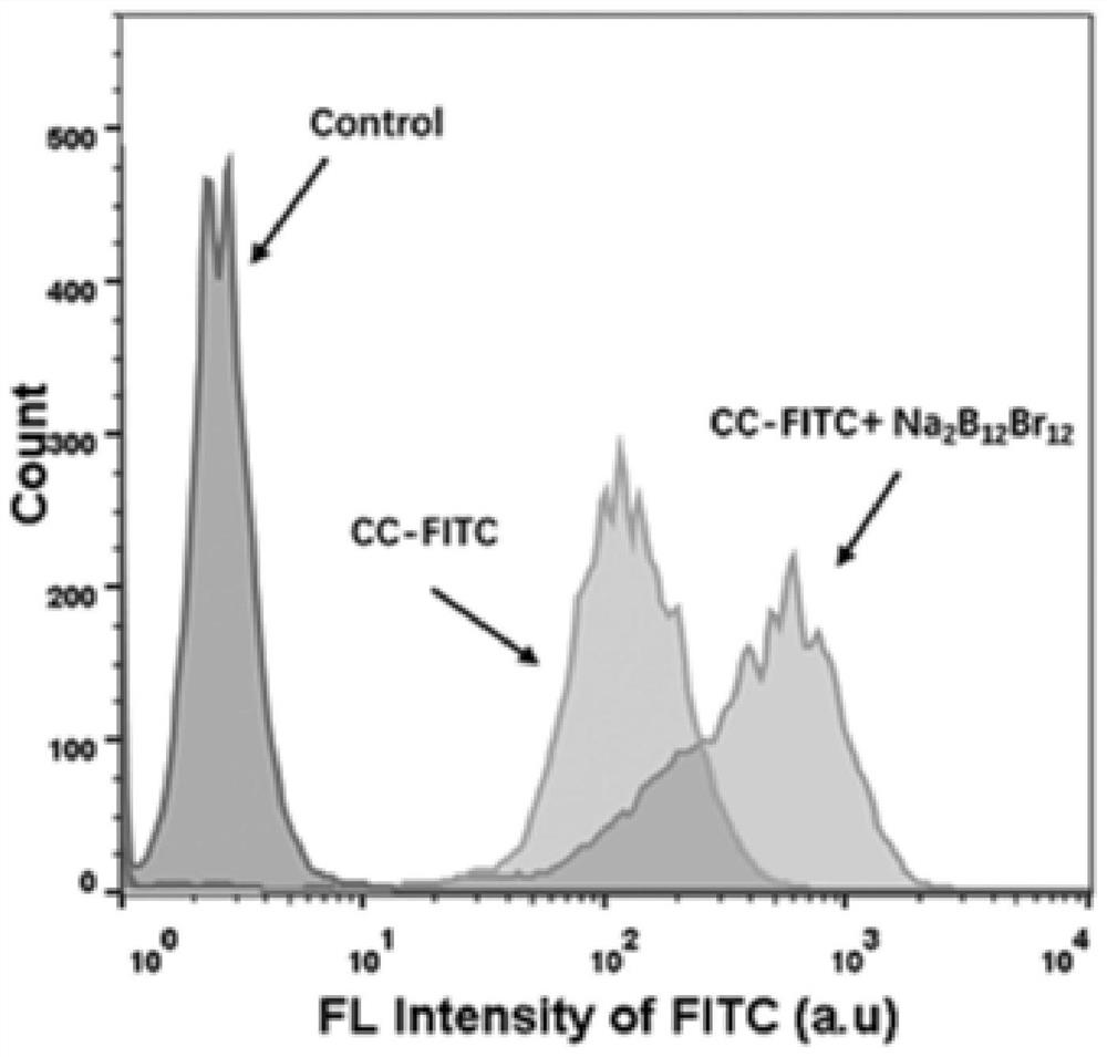
[0036] (4) Use a confocal microscope to perform confocal imaging of the cells to observe the distribution of the labeled cytochrome C in the cells. The fluorescent channel of the labeled cytochrome C at 630-670nm can be used to see the distribution of proteins in the cells. Flow cytometry The instrument detects the efficiency of labeled cytochro...


PUM


Abstract

The invention discloses a method for intracellular delivery of protein. The method comprises the following steps of (1) preparing a protein solution and a halogenatedboron cluster solution with certain concentrations; (2) slowly adding the halogenatedboron cluster solution into the protein solution, quickly and uniformly mixing the two solutions, and standing for a period of time; and (3) adding amixed solution of halogenatedboron clusters and protein into a standard complete culture medium, and co-culturing the mixed solution with to-be-transferred cells for a period of time. The halogenatedboron clusters can be used to deliver proteins across biofilms without modification by chemical bonds. The method for protein transferring can prevent capture by endosomes, is simple and easy to implement, low in cost and relatively high in transfer efficiency, and the halogenatedboron clusters are a protein transfer carrier with a wide prospect.

Description

technical field [0001] The invention relates to the technical field of bioengineering, in particular to a method for intracellular protein delivery. Background technique [0002] Protein is one of the material basis of life and the main bearer of life activities. Research at this stage has found that many diseases are related to the loss of normal proteins in cells. Transporting proteins across cell membranes can introduce missing proteins caused by genetic defects and therapeutic proteins into cells for the treatment of certain diseases, such as respiratory diseases, cancer, arthritis, and more. Protein drugs have the advantages of low toxicity, strong specificity, and clear functions, but they have disadvantages such as poor stability, high clearance rate, and poor transmembrane ability, which have caused great obstacles to the clinical application of proteins. [0003] In order to improve the bioavailability and therapeutic effect of proteins, a series of carriers for i...

Claims


Application Information

Patent Timeline
Patent Type & Authority Applications(China)
IPC IPC(8): C12N5/00
CPCC12N5/00C12N2500/05C12N2501/998
Inventor 王晓娟张智雄马锡琦维尔纳·M·诺黄方戴祺
Owner CHINA UNIV OF PETROLEUM (EAST CHINA)
Why Eureka
Try Eureka