Breast nodule detection method and system based on ultrasonic medicine

A breast nodule and detection method technology, applied in medical simulation, medical imaging, medical informatics, etc., can solve the problems of dynamic recognition and identification of breast tumors, and achieve the effect of solving the difficulties of dynamic identification and identification and the stability of pre-training

Active Publication Date: 2020-12-15
厦门大学附属翔安医院
View PDF5 Cites 8 Cited by
  • Summary
  • Abstract
  • Description
  • Claims
  • Application Information

AI Technical Summary

Problems solved by technology

[0004] The present invention proposes a breast nodule detection method and system based on ultrasound medicine, using ultrasound breast video image inspection as the main means, aiming to establish an accurate method for judging benign and malignant breast tumors and pathological typing, and solving the problem of dynamic identification and differentiation of breast tumors Difficult clinical problems

Method used


Image

  • Breast nodule detection method and system based on ultrasonic medicine
  • Breast nodule detection method and system based on ultrasonic medicine
  • Breast nodule detection method and system based on ultrasonic medicine

Examples


Embodiment 1

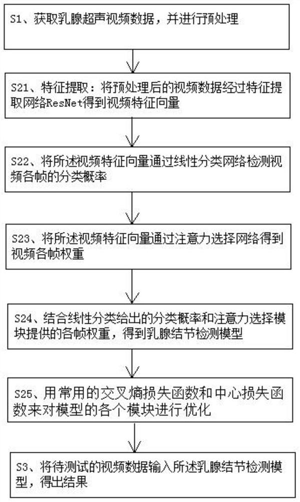
[0044] Cooperate figure 1 As shown, the present invention discloses a breast nodule detection method based on ultrasound medicine, comprising the following steps:

[0045] S1. Obtain breast ultrasound video data and perform preprocessing;

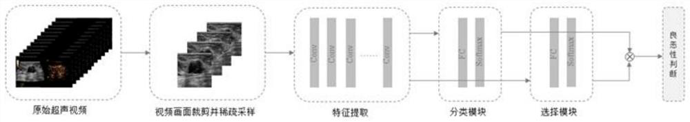
[0046] S11. Intercept the largest rectangle from the breast ultrasound original video data at the ultrasound imaging site, and uniformly scale the size to 256×256, wherein 20 frames are equally spaced and sparsely sampled in the middle of the video;

[0047] S12. Digital image processing, classifying the intercepted ultrasound video images, selecting clearer image slices containing complete nodules, and deleting redundant images that do not contain regions of interest;

[0048] S13. Data enhancement. The classified ultrasound video images are randomly cropped into 224×224 pixels by random cropping, and one ultrasound video image can be enhanced several hundred times. The spatial geometric transformation is used to affine the ultrasound video...

Embodiment 2

[0072] The breast nodule detection system based on ultrasonic medicine of the present invention is characterized in that it includes a feature extraction module, a linear classification module, an attention selection module, a video detection module and a loss function module, wherein:

[0073] Feature extraction module: the preprocessed video data is obtained through the feature extraction network ResNet to obtain the video feature vector;

[0074] Linear classification module: the classification probability of each frame of video is detected by the video feature vector through a linear classification network;

[0075] Attention selection module: the video feature vector is passed through the attention selection network to obtain the weight of each frame of the video;

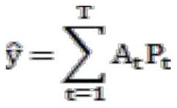
[0076] Video detection module: Combining the classification probability given by linear classification and the weight of each frame provided by the attention selection module, a breast nodule detection model i...


PUM


Abstract

The invention discloses a breast nodule detection method and system based on ultrasonic medicine. The method comprises the steps of obtaining breast ultrasonic video data, carrying out the preprocessing, carrying out training on the preprocessed data through a transfer learning method, building a breast nodule detection model, inputting to-be-tested video data into the breast nodule detection model, and obtaining a result. The breast nodule detection method and system based on ultrasonic medicine aim to establish a precise method for judging benign and malignant breast tumors and carrying outpathological typing by taking ultrasonic breast video image examination as a main means, and solve the difficult clinical problem that dynamic identification and identification of breast tumors are difficult. The establishment of the highly accurate breast tumor qualitative system is of great clinical significance to the diagnosis and treatment of breast tumor patients.

Description

technical field [0001] The invention relates to the technical field of video image processing, in particular to a breast nodule detection method and system based on ultrasound medicine. Background technique [0002] Ultrasound computer-aided computer-aided diagnosis (Computer Aided Diagnosis, CAD) can detect the internal information of tumors that cannot be obtained by human eyes, such as texture information, edge information, etc., and provide reliable auxiliary diagnosis opinions for doctors. It reduces the misdiagnosis caused by doctors' inexperience or visual fatigue, and also reduces the biopsy rate and pain for patients. The artificial intelligence real-time detection module of breast ultrasound used in the study can automatically help doctors find nodules during ultrasound exploration, which is of great significance for reducing missed diagnoses and can help doctors self-train ultrasound diagnosis capabilities. The combination of breast ultrasound artificial intellig...

Claims


Application Information

Patent Timeline
Patent Type & Authority Applications(China)
IPC IPC(8): G16H50/50G16H30/40G06T7/11G06T7/00G06T3/00G06K9/62G06K9/00
CPCG16H50/50G16H30/40G06T7/0012G06T3/0006G06T7/11G06T2207/30096G06T2207/30068G06V20/41G06F18/214
Inventor 张国君李卫斌陈敏王连生陈云超徐辉雄
Owner 厦门大学附属翔安医院
Why Eureka
Try Eureka