Broken bone segmentation method and device for three-dimensional image, computer equipment and storage medium

A technology of three-dimensional image and bone crushing, which is applied in the field of intelligent medical treatment, can solve the problem that the segmentation method of bone crushing cannot consider the correlation of tissue space, etc., and achieve the effect of improving accuracy and efficiency and saving segmentation time

Active Publication Date: 2021-01-19
PING AN TECH (SHENZHEN) CO LTD
View PDF7 Cites 2 Cited by
  • Summary
  • Abstract
  • Description
  • Claims
  • Application Information

AI Technical Summary

Problems solved by technology

[0003] Aiming at the problem that the existing bone fragment segmentation method cannot consider the spatial correlation of tissue, a method, device, computer equipment and storage medium for bone fragment segmentation aiming at three-dimensional images that can consider the spatial correlation of tissue are provided

Method used


Image

  • Broken bone segmentation method and device for three-dimensional image, computer equipment and storage medium
  • Broken bone segmentation method and device for three-dimensional image, computer equipment and storage medium
  • Broken bone segmentation method and device for three-dimensional image, computer equipment and storage medium

Examples


Embodiment 1

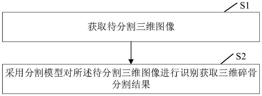
[0059] see figure 1, a bone fragment segmentation method of a three-dimensional image of the present embodiment, comprising:

[0060] S1. Acquire a three-dimensional image to be segmented.

[0061] It should be noted that the three-dimensional image to be segmented is a three-dimensional MRI (Magnetic Resonance Imaging, nuclear magnetic resonance imaging) image. MRI displays the internal information of the body in the form of images, and has the advantages of non-invasive, multi-modal, and accurate positioning.

[0062] S2. Using a segmentation model to identify the 3D image to be segmented to obtain a 3D bone fragment segmentation result.

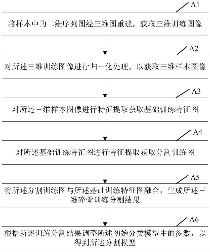
[0063] Wherein, the segmentation model includes a feature extraction module, an intermediate module and a segmentation module.

[0064] Specifically, step S2 uses a segmentation model to identify the 3D image to be segmented to obtain a 3D bone fragment segmentation result including:

[0065] S21. Perform feature extraction on the 3D i...

Embodiment 2

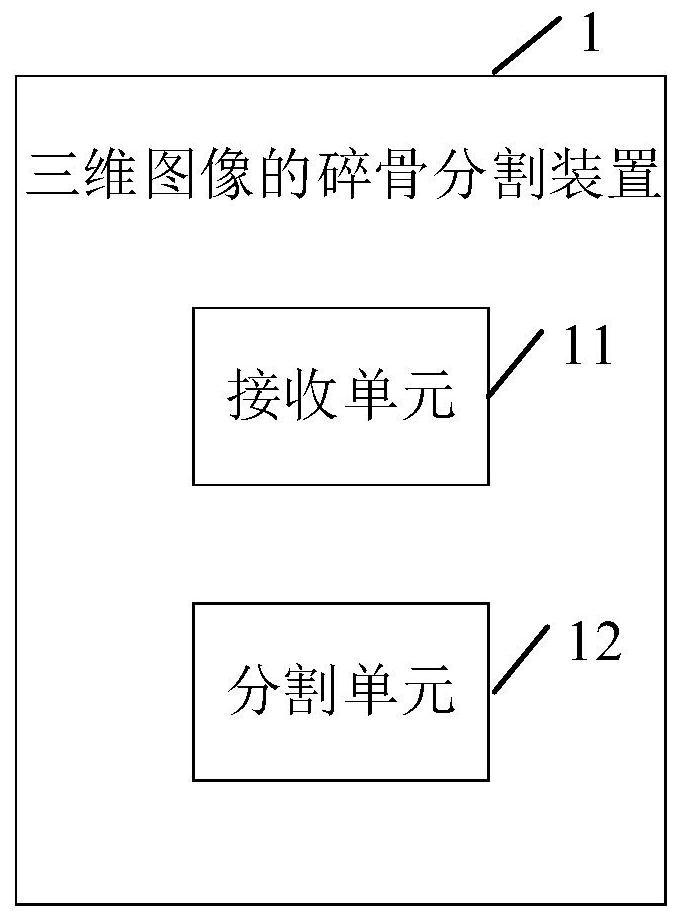
[0120] see image 3 , a three-dimensional image bone segmentation device 1 in this embodiment includes: a receiving unit 11 and a segmentation unit 12 .

[0121] The receiving unit 11 is configured to acquire a three-dimensional image to be segmented.

[0122] It should be noted that the three-dimensional image to be segmented is a three-dimensional MRI image. MRI displays the internal information of the body in the form of images, and has the advantages of non-invasive, multi-modal, and accurate positioning.

[0123] The segmentation unit 12 is configured to use a segmentation model to identify the 3D image to be segmented to obtain a 3D bone fragment segmentation result.

[0124] Among them, see Figure 4 The segmentation model shown includes a feature extraction module 121 , an intermediate module 122 and a segmentation module 123 .

[0125] The segmentation unit 12 performs feature extraction on the three-dimensional image to be segmented to obtain a basic feature map ...

Embodiment 3

[0141] In order to achieve the above object, the present invention also provides a computer device 2, the computer device 2 includes a plurality of computer devices 2, the components of the three-dimensional image bone segmentation device 1 in the second embodiment can be dispersed in different computer devices 2 , the computer device 2 can be a smart phone, a tablet computer, a notebook computer, a desktop computer, a rack server, a blade server, a tower server or a cabinet server (including an independent server, or a combination of multiple servers) for executing programs server cluster), etc. The computer equipment 2 of the present embodiment at least includes but is not limited to: a memory 21, a processor 23, a network interface 22 and a three-dimensional image bone segmentation device 1 that can communicate with each other through a system bus (refer to Figure 5 ). It should be pointed out that, Figure 5 Only the computer device 2 is shown with components - but it s...


PUM


Abstract

The invention discloses a broken bone segmentation method and device for a three-dimensional image, computer equipment and a storage medium, and belongs to the field of intelligent medical treatment.According to the broken bone segmentation method for the three-dimensional image, the three-dimensional image serves as input, feature extraction is conducted on the three-dimensional image to be segmented through a feature extraction module in a segmentation model to obtain a basic feature map, and the correlation of tissues in the three-dimensional image is considered; the basic feature map is subjected to feature extraction through the middle module to obtain the segmentation map with the same size as the original to-be-segmented three-dimensional image, the segmentation map and the basic feature map are fused through the segmentation module, then the three-dimensional broken bone segmentation result is obtained, the segmentation time is saved, and the segmentation precision and efficiency are improved.

Description

technical field [0001] The invention relates to the field of intelligent medical treatment, in particular to a bone fragment segmentation method, device, computer equipment and storage medium of a three-dimensional image. Background technique [0002] Fractures are a common disease. More severe fractures often have broken bones. If broken bones are missed during surgery, serious consequences may result. The existing traditional segmentation method is mainly to understand the image through artificial subjective consciousness, so as to extract specific feature information, such as: gray level information, texture information and symmetry information, etc. to realize the segmentation of bone fragments. However, this segmentation method is only suitable for specific images. Good segmentation results, but there are problems that the segmentation results are too rough and the segmentation efficiency is low. With the emergence of artificial intelligence, especially deep learning, ...

Claims


Application Information

Patent Timeline
Patent Type & Authority Applications(China)
IPC IPC(8): G06T7/00G06T7/10G06T17/00G06K9/46G06N3/04G06N3/08
CPCG06T7/0012G06T7/10G06T17/00G06T2207/10088G06T2207/30008
Inventor 洪振厚王健宗瞿晓阳
Owner PING AN TECH (SHENZHEN) CO LTD
Why Eureka
Try Eureka