Medical image lesion segmentation method

A medical image and lesion technology, applied in the field of medical image lesion segmentation, can solve problems such as low segmentation accuracy, gradient disappearance, and weak learning ability, and achieve the effects of strong reproducibility, reduced feature loss, and reduced medical costs

Inactive Publication Date: 2021-01-22
山西三友和智慧信息技术股份有限公司
View PDF5 Cites 14 Cited by
  • Summary
  • Abstract
  • Description
  • Claims
  • Application Information

AI Technical Summary

Problems solved by technology

[0003] Reasons for problems or defects: 1. Due to the complex types of hospital equipment and various data, there is no unified standard to standardize the data to be collected, and there is no unified medical database; secondly, the data ratio of medical images is unbalanced, The ratio of disease data to normal data fluctuates greatly, and is directly related to the type of disease; finally, specific doctors are required to provide technical guidance for different diseased parts, and different doctors have different guidance opinions, so there is no way to form a unified standard
2. Traditional segmentation methods often require a large number of human-computer interaction processes to complete target extraction, and the learning ability is weak, and the resistance to interference such as noise and blur is weak, resulting in low segmentation accuracy
3. The current Unet network is difficult to fully extract medical image features. At the same time, there are problems such as gradient disappearance and low feature utilization during the training process, resulting in low segmentation accuracy of the model.

Method used


Image

  • Medical image lesion segmentation method
  • Medical image lesion segmentation method
  • Medical image lesion segmentation method

Examples


Embodiment Construction

[0037] The following will clearly and completely describe the technical solutions in the embodiments of the present invention with reference to the accompanying drawings in the embodiments of the present invention. Obviously, the described embodiments are only some, not all, embodiments of the present invention. Based on the embodiments of the present invention, all other embodiments obtained by persons of ordinary skill in the art without making creative efforts belong to the protection scope of the present invention.

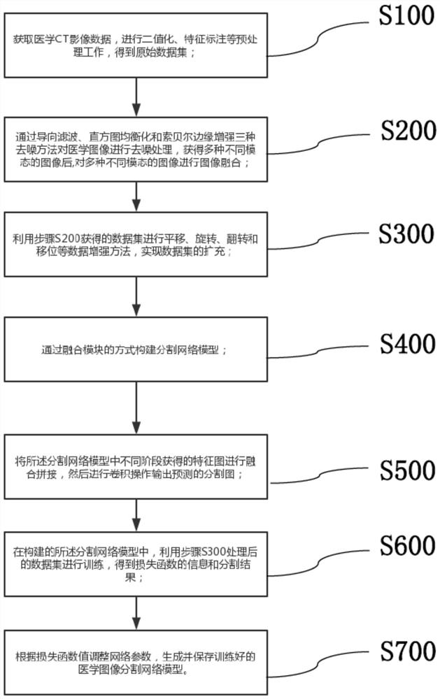
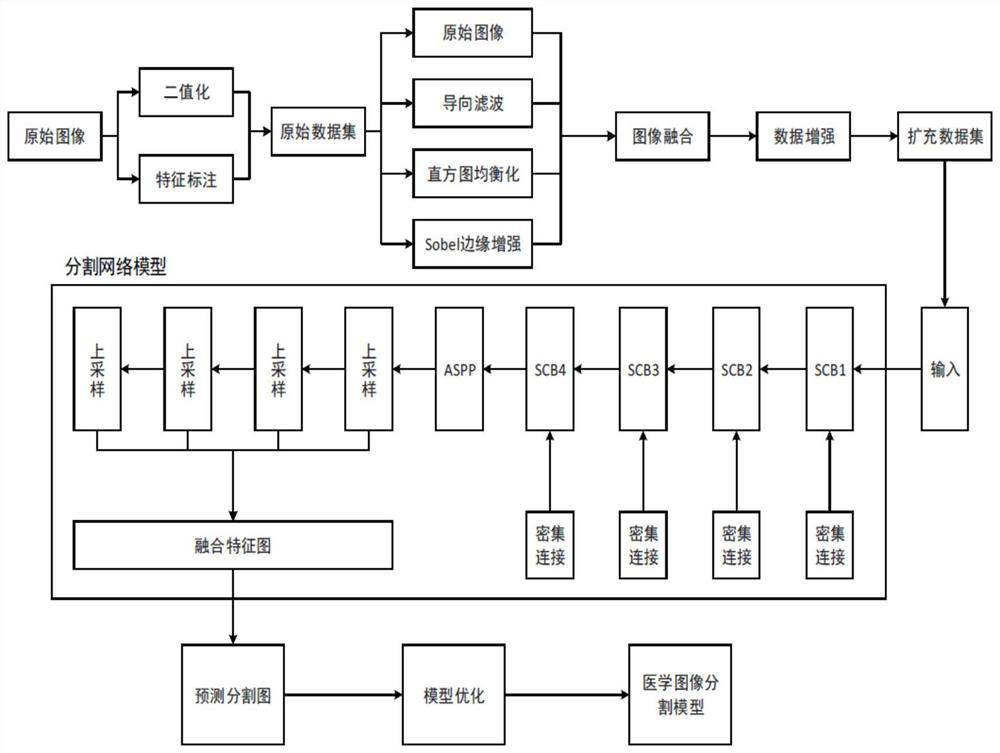
[0038] A medical image lesion segmentation method, such as figure 1 shown, including the following steps:

[0039] S100. Acquire medical CT image data, perform preprocessing such as binarization and feature labeling, and obtain an original data set;

[0040] S200, use three denoising methods of guided filtering, adaptive histogram equalization and Sobel Sobel edge enhancement to denoise the original medical image respectively, obtain images of four different ...


PUM


Abstract

The invention belongs to the technical field of artificial intelligence image processing, and particularly relates to a medical image lesion segmentation method, which comprises the following steps ofobtaining medical CT image data, and performing preprocessing work such as binarization and feature labeling to obtain an original data set; carrying out denoising processing on the medical image byusing a denoising method, and carrying out image fusion on various images with different modes; carrying out data enhancement methods such as translation, rotation, overturning and shifting by utilizing the obtained data set to realize expansion of the data set; constructing a segmentation network model through a fusion module; fusing and splicing the feature maps obtained in different stages in the segmentation network model, and performing convolution operation to output a predicted segmentation map; in the constructed segmentation network model, carrying out training by using the data set to obtain information of a loss function and a segmentation result; and according to the medical image segmentation method and system, generating and storing a trained medical image segmentation network model. The segmentation accuracy of the model is improved.

Description

technical field [0001] The invention belongs to the technical field of artificial intelligence image processing, and in particular relates to a method for segmenting medical image lesions. Background technique [0002] With the advancement of computer science and technology and the improvement of computer hardware performance, imaging technology is also developing continuously, and medical imaging, as a discipline with rapid development in the direction of medicine, has gradually become a research hotspot. However, due to the differences in the imaging principles of medical images and the characteristics of human tissues, medical images are characterized by complexity and diversity, and it is difficult to identify and segment them. At the same time, the development of medical images provides people with rich image data information, making doctors It takes a lot of time to identify symptoms, which will be a challenge for doctors, and may cause diagnostic fatigue due to lack o...

Claims


Application Information

Patent Timeline
Patent Type & Authority Applications(China)
IPC IPC(8): G06T7/00G06T5/00G06T5/20G06T5/50G06T7/11G06N3/04
CPCG06T7/0012G06T5/002G06T5/50G06T7/11G06T5/20G06T2207/10081G06T2207/20221G06T2207/20081G06T2207/20084G06N3/045
Inventor 潘晓光张海轩王小华令狐彬张娜焦璐璐
Owner 山西三友和智慧信息技术股份有限公司
Why Eureka
Try Eureka