
Parallel imaging probe based on fine spectral coding of virtual image phased array

A phased array and imaging probe technology, which is applied in the field of optical endoscopy to achieve the effects of simple design, improved field of view, and reduced speckle noise.

Active Publication Date: 2021-03-16
ZHEJIANG UNIV
View PDF6 Cites 2 Cited by
  • Summary
  • Abstract
  • Description
  • Claims
  • Application Information

AI Technical Summary

Problems solved by technology

Unlike diffraction gratings, VIPA uses the principle of multi-beam interference and has high spectral resolution, but its free spectral range (FSR) is very narrow.

Method used


Image

  • Parallel imaging probe based on fine spectral coding of virtual image phased array
  • Parallel imaging probe based on fine spectral coding of virtual image phased array
  • Parallel imaging probe based on fine spectral coding of virtual image phased array

Examples


Embodiment Construction

[0043] The present invention will be described in detail below in conjunction with the accompanying drawings and embodiments, but the present invention is not limited thereto.

[0044] A clinical endoscopic probe that meets the following technical indicators:

[0045] 1. The working wavelength covers the visible light range of 400-800nm, and the central wavelength is 550-650nm;

[0046] 2. The diameter of the bare probe (before packaging) is less than 1mm;

[0047] 3. The length of the hard end of the probe is less than 7mm;

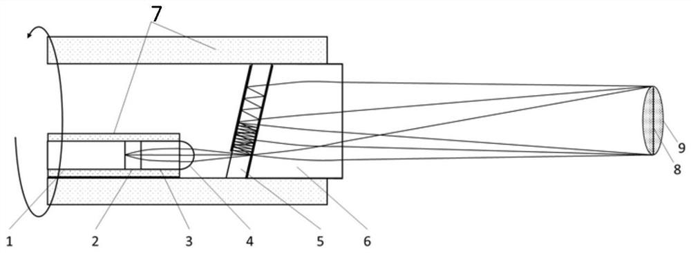
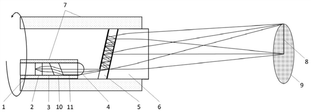
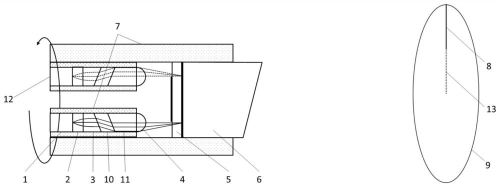
[0048] 4. The number of effective pixels in the linear field of view 8 of the probe is greater than 200, or the number of pixels in the circular field of view 9 is greater than 160,000;

[0049] 5. The field of view or FAR of the probe is greater than 20 degrees;

[0050] The schematic diagram of the probe is as figure 1 , Figure 4 shown. Among them, SMF1 is the 630-HP optical fiber of Nufern Company, which is suitable for transmitting visible lig...


PUM


Abstract

The invention discloses a parallel imaging probe based on fine spectral coding of a virtual image phased array. The parallel imaging probe is composed of a rotary illumination probe and a fixed collection channel, the illumination probe uses a VIPA with high spectral resolution and a narrow free spectral range as a dispersion element to perform spatial coding on an emergent light beam, and a spectral comb with different spectral fine structures but with a spectral range still equal to that of incident white light is formed in the one-dimensional direction of the sample space. The illuminationprobe is driven by an external motor to rotate around the axis of the probe, so that a two-dimensional circular view field is formed. Light scattered or reflected from the sample is conducted by the fixed collection channel to a fine spectral detection device. According to the spatial coding based on the VIPA, parallel imaging in the one-dimensional direction can be achieved, the fine spectrum ofthe spatial coding still keeps the wide spectrum characteristic of the original incident white light, color imaging of an optical endoscope can be ensured, and therefore great clinical application potential is achieved.

Description

technical field [0001] The invention belongs to the field of optical endoscopy, in particular to a parallel imaging probe using a virtual image phase array (VIPA) for fine spectrum encoding. [0002] technical background [0003] Endoscopy provides doctors with intuitive optical images of the inside of the human body. It has the characteristics of low damage and high resolution, so it is widely used to observe and diagnose lesions in tissues or organs such as the digestive tract, reproductive tract, and respiratory tract. The earliest endoscope can be traced back to 60 years ago [1]. Hirschowitz et al. developed a clinical endoscope model using a coherent fiber bundle (CFB) and performed an examination of the upper gastrointestinal tract. This kind of endoscope, which uses CFB light guide at the far end and charge-coupled device (CCD) or complementary metal oxide semiconductor (complementary metal oxide semiconductor (CMOS)) detection at the proximal end, has a certain degree...

Claims


Application Information

Patent Timeline
Patent Type & Authority Applications(China)
IPC IPC(8): A61B1/00A61B1/05A61B1/07
CPCA61B1/00163A61B1/05A61B1/07A61B1/00096A61B1/00172A61B1/0605
Inventor 丁志华章程邱建榕
Owner ZHEJIANG UNIV
Learn More