
Microscopic shooting method for joint development of single-refraction plant tissue and double-refraction plant tissue

A technology of plant tissue and birefringence, applied in microscopes, optics, instruments, etc., to achieve rapid resolution, easy popularization, and rapid inspection

Pending Publication Date: 2021-04-02
崔亚君
View PDF3 Cites 0 Cited by
  • Summary
  • Abstract
  • Description
  • Claims
  • Application Information

AI Technical Summary

Problems solved by technology

[0004] The purpose of the present invention is to provide a microscopic shooting method of single and double refraction plant tissue imaging to solve the deficiencies of the prior art

Method used


Image

  • Microscopic shooting method for joint development of single-refraction plant tissue and double-refraction plant tissue
  • Microscopic shooting method for joint development of single-refraction plant tissue and double-refraction plant tissue
  • Microscopic shooting method for joint development of single-refraction plant tissue and double-refraction plant tissue

Examples


Embodiment 1

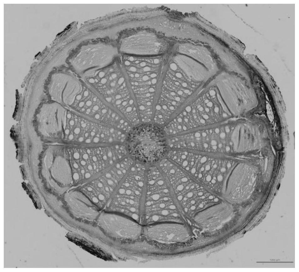
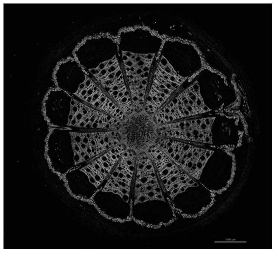
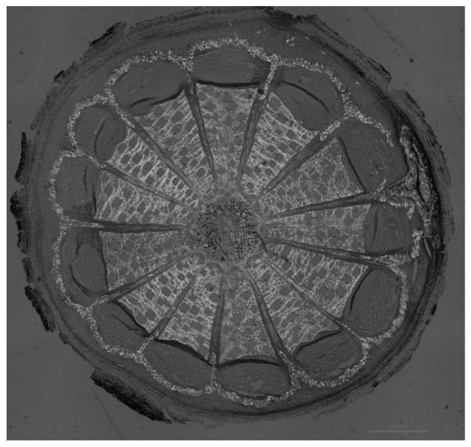
[0028] (1) Take the cane of Akebia chinensis, soften it, and make thin slices of 10-20 μm by freehand, slide-away sectioning or permanent paraffin embedding. Except for direct observation of permanent paraffin sections, select flat slices for other sections and put them on glass slides, add 1-2 drops of chloral hydrate test solution, heat permeabilization on an alcohol lamp, and then add 1-2 drops of dilute glycerin, Cover with a coverslip.

[0029] (2) Under a polarized light microscope, the cane of the said Bai Mutong is observed, and the polarizer and the analyzer are rotated 17° to the left or right from the orthogonal state, and the observation is made under a normal bright field. Microscopic features of monorefringent tissue and microscopic features of birefringent tissue under cross-polarized light.

[0030] (3) Using the large-scale image stitching technology of the digital imaging system and the real-time depth-of-field imaging technology to shoot the images under th...

Embodiment 2

[0035] (1) Take the cane of Akebia chinensis, soften it, and make thin slices of 10-20 μm by freehand, slide-away sectioning or permanent paraffin embedding. Except for direct observation of permanent paraffin sections, select flat slices for other sections and put them on glass slides, add 1-2 drops of chloral hydrate test solution, heat permeabilization on an alcohol lamp, and then add 1-2 drops of dilute glycerin, Cover with a coverslip.

[0036] (2) if Figure 5 As shown, place the microplate under a polarized light microscope, insert a λ plate optical path compensator in the slot of the middle lens barrel located at 45 degrees between the polarizer and the analyzer, Observing the microscopic characteristics of single refraction tissue under normal bright field and the microscopic characteristics of birefringence tissue under orthogonal polarized light under the condition of 90° orthogonal;

[0037] (3) Using the large-scale image stitching technology of the digital imag...


PUM


Abstract

The invention relates to the field of polarized light detection, in particular to a microscopic shooting method for the joint development of a single-refraction plant tissue and a double-refraction plant tissue. The method comprises the following steps: (1) taking a to-be-observed part of a plant tissue, and preparing a plant tissue slide according to a microscopic slide preparation method under four microscopic identification methods (general rule 2001) of Chinese Pharmacopoeia in edition 2020; (2) taking the slide in the step (1), placing the slide under a polarized light microscope, and adjusting a basic imaging mechanism to be in single-refraction and double-refraction tissue structure common imaging mode; and (3) shooting an image by adopting a large-image splicing technology and a real-time depth-of-field extension imaging technology of a digital imaging system. According to the method, the defects that partial tissue structure characteristic information of a single-refraction microscopic image is lost under the traditional orthogonal polarized light condition, the microscopic image cannot quickly and accurately position a double-refraction microscopic characteristic marker under the normal light condition, and the definition and the integrity of the image are difficult to consider at the same time during microscopic shooting imaging are overcome.

Description

technical field [0001] The invention relates to the field of polarized light detection, in particular to a microphotography method for co-imaging single and double refraction plant tissues. Background technique [0002] At present, authoritative documents and quality standards for microscopic identification in the field of microscopic observation and imaging of plants, especially in the field of identification of crude drugs (including traditional Chinese medicines), have relatively detailed descriptions of microscopic characteristics of most crude drugs (including traditional Chinese medicines), but with The supporting illustrations are limited by relevant equipment and technical methods, and the presentation forms are all powder feature maps taken in a single shot, mosaic maps of discontinuous local features of plant sections taken under the normal bright field of conventional microscopes or hand-painted ink line maps, as well as images using recent years. The polarized li...

Claims


Application Information

Patent Timeline
Patent Type & Authority Applications(China)
IPC IPC(8): G02B21/00G02B21/36
CPCG02B21/0092G02B21/361
Inventor 崔亚君马颖娴
Owner 崔亚君
Learn More