Medical image processing method, device and equipment and storage medium

A technology of medical image and processing method, applied in the field of image processing, which can solve problems such as reducing noise level

Pending Publication Date: 2021-04-06
SHENZHEN INST OF ADVANCED TECH
View PDF0 Cites 0 Cited by
  • Summary
  • Abstract
  • Description
  • Claims
  • Application Information

AI Technical Summary

Problems solved by technology

[0004] The embodiment of the present invention provides a medical image processing method, device, equipment and storage medium, which sol

Method used


Image

  • Medical image processing method, device and equipment and storage medium
  • Medical image processing method, device and equipment and storage medium
  • Medical image processing method, device and equipment and storage medium

Examples


Embodiment 1

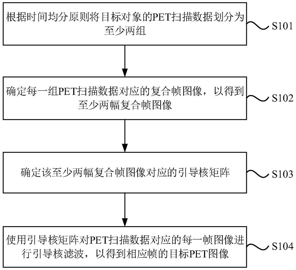
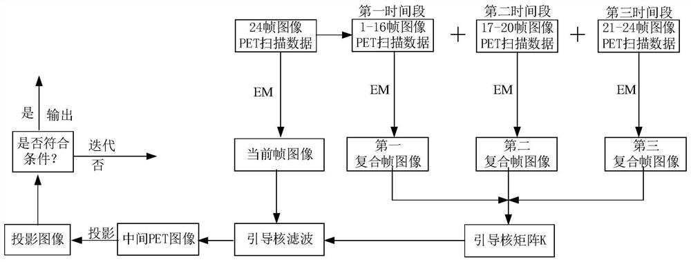
[0028] figure 1 It is a flow chart of the medical image processing method provided by Embodiment 1 of the present invention. The technical solution of this embodiment is suitable for determining the guiding kernel matrix carrying spatial information through at least two composite frame images, so that when the guiding kernel matrix filters each frame of PET image data, it also filters the corresponding frame image Supplementing spatial information so that the target PET image generated by filtering has both a low noise level and rich spatial information. The method can be executed by the medical image processing apparatus provided in the embodiment of the present invention, and the apparatus can be implemented in the form of software and / or hardware, and configured to be applied in a processor of an image processing device. The method specifically includes the following steps:

[0029] S101. Divide the PET scan data of the target object into at least two groups according to ...

Embodiment 2

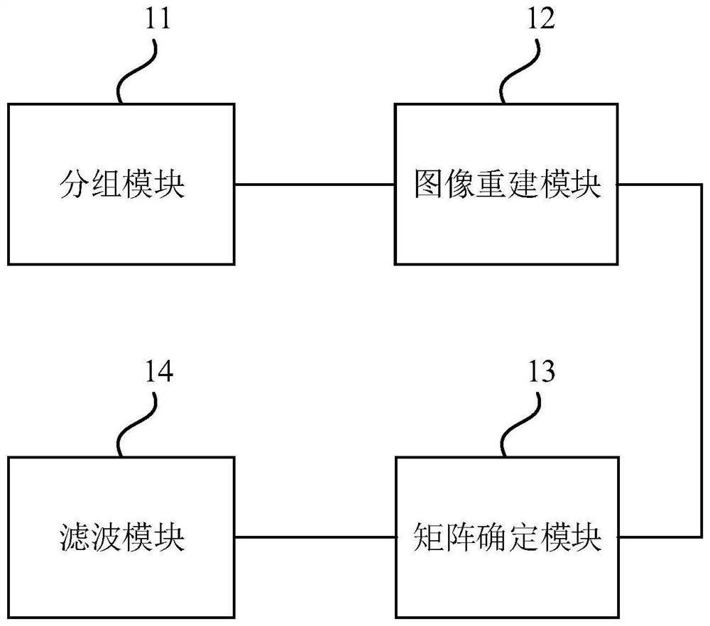
[0055] image 3 It is a structural block diagram of a medical image processing device provided by an embodiment of the present invention. The device is used to execute the medical image processing method provided in any of the above embodiments, and the device may be implemented by software or hardware. The unit includes:

[0056] The grouping module 11 is used for dividing the PET scan data of the target object into at least two groups according to the principle of equal time division;

[0057] The image reconstruction module 12 is used to determine the composite frame images corresponding to each group of PET scan data, so as to obtain at least two composite frame images;

[0058] A matrix determination module 13, configured to determine the guiding kernel matrix corresponding to the at least two composite frame images;

[0059] The filtering module 14 is configured to perform guided kernel filtering on each frame of image corresponding to the PET scan data by using the g...

Embodiment 3

[0068] Figure 4 A schematic structural diagram of an image processing device provided in Embodiment 3 of the present invention, such as Figure 4 As shown, the device includes a processor 201, a memory 202, an input device 203, and an output device 204; the number of processors 201 in the device may be one or more, Figure 4 Take a processor 201 as an example; the processor 201, memory 202, input device 203, and output device 204 in the device can be connected by bus or other methods, Figure 4 Take connection via bus as an example.

[0069] The memory 202, as a computer-readable storage medium, can be used to store software programs, computer-executable programs and modules, such as program instructions / modules corresponding to the medical image processing method in the embodiment of the present invention (for example, grouping module 11, image reconstruction module 12, matrix determination module 13 and filtering module 14). The processor 201 executes various functional ...


PUM


Abstract

The embodiment of the invention discloses a medical image processing method, device and equipment and a storage medium. The method comprises the steps of dividing PET scanning data of a target object into at least two groups according to a time sharing principle; determining a composite frame image corresponding to each group of PET scanning data to obtain at least two composite frame images; determining guide kernel matrixes corresponding to the at least two composite frame images; and performing guided kernel filtering on each frame of image corresponding to the PET scanning data by using the guided kernel matrix to obtain a target PET image of the corresponding frame. The problem that the noise level of the PET image cannot be reduced under the condition of not influencing the space information carried by the PET image in the prior art is solved.

Description

technical field [0001] Embodiments of the present invention relate to the field of image processing, and in particular, to a medical image processing method, device, equipment, and storage medium. Background technique [0002] Positron Emission Computed Tomography (PET) is a functional imaging instrument and a relatively advanced clinical imaging technology in the field of nuclear medicine. PET imaging technology is currently the only technology that uses anatomical forms for functional, metabolic and receptor imaging. It is non-invasive and can provide three-dimensional and functional images of the whole body. Before performing PET imaging on patients, radiopharmaceuticals (tracers) with short half-lives need to be injected into patients. Since the distribution of tracers changes rapidly, patients must be scanned at a high scanning speed. The higher scanning speed is prone to undercounting when the dose of tracer is low, and undercounting will reduce the image quality of P...

Claims


Application Information

Patent Timeline
IPC IPC(8): G06T11/00G06T5/00
CPCG06T5/002G06T11/006G06T11/008
Inventor 郑海荣刘新张娜胡战利杨永峰梁栋欧阳张磊
Owner SHENZHEN INST OF ADVANCED TECH
Why Eureka
Try Eureka