
In-situ 3D printing cartilage repair device and method

A cartilage repair and 3D printing technology, which is applied in the field of additive manufacturing, can solve problems such as the inability to meet the complex environment and the inability to guarantee the accuracy of repair, and achieve the effects of short manual intervention time, fast printing speed, and easy storage

Pending Publication Date: 2021-04-20
SOUTH CHINA UNIV OF TECH +1
View PDF0 Cites 0 Cited by
  • Summary
  • Abstract
  • Description
  • Claims
  • Application Information

AI Technical Summary

Problems solved by technology

However, this method of holding a biological gun by a doctor cannot guarantee the repair accuracy requirements, and can only print and repair cartilage defects with simple shapes, which cannot meet the complex environment in the human body.

Method used


Image

  • In-situ 3D printing cartilage repair device and method
  • In-situ 3D printing cartilage repair device and method
  • In-situ 3D printing cartilage repair device and method

Examples


Embodiment

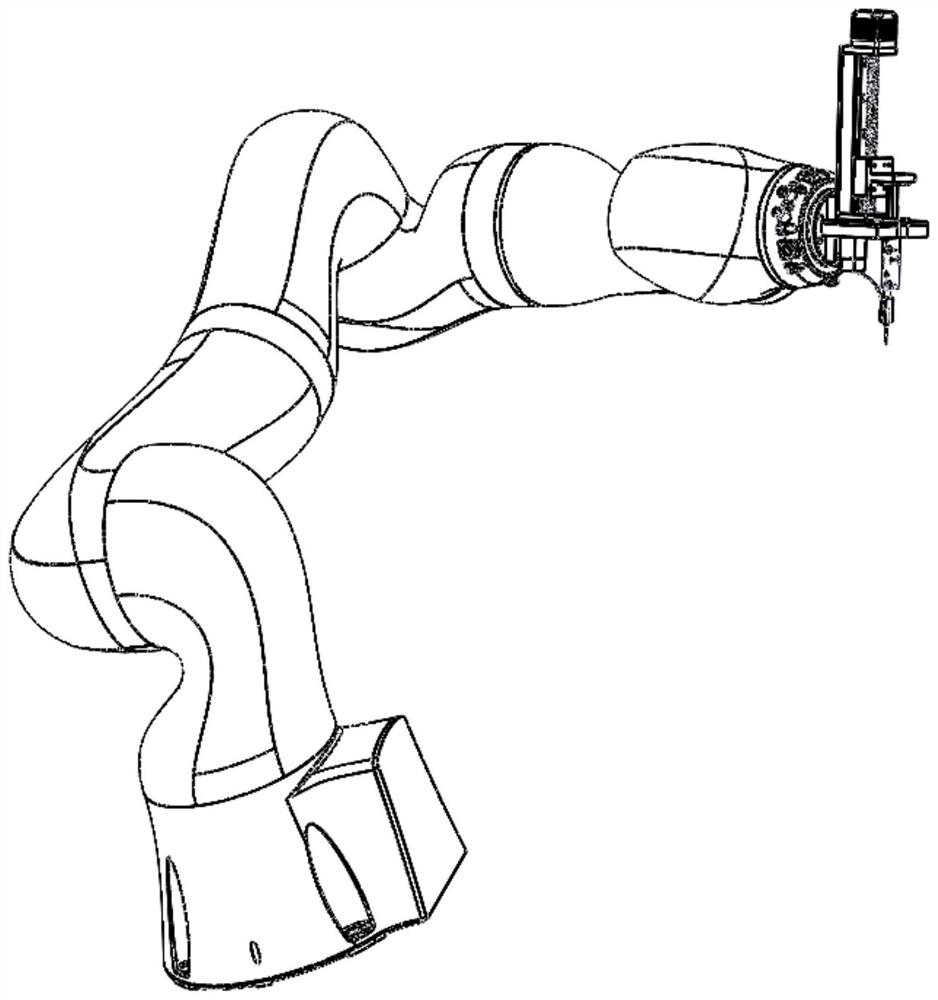
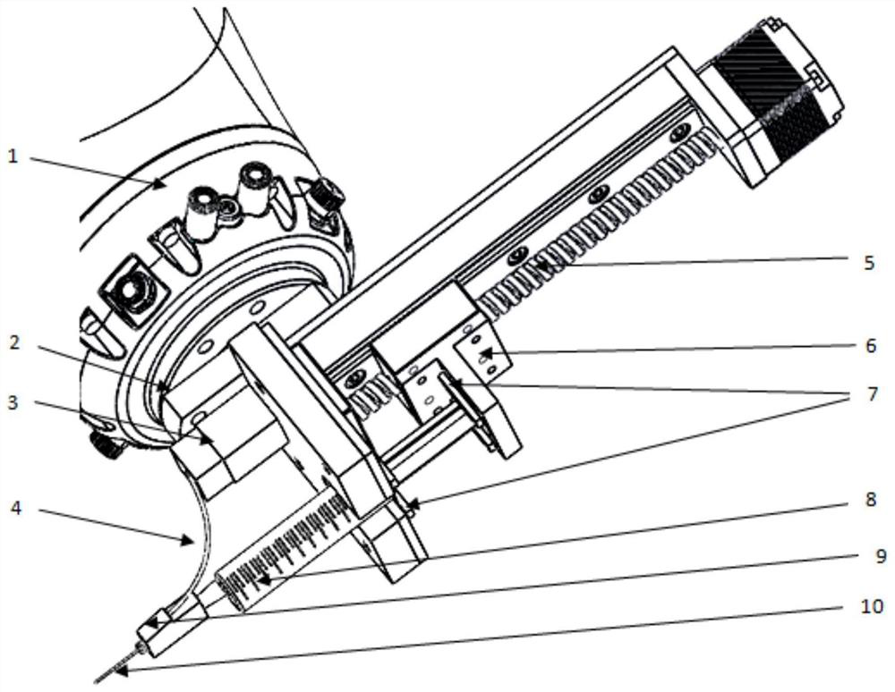
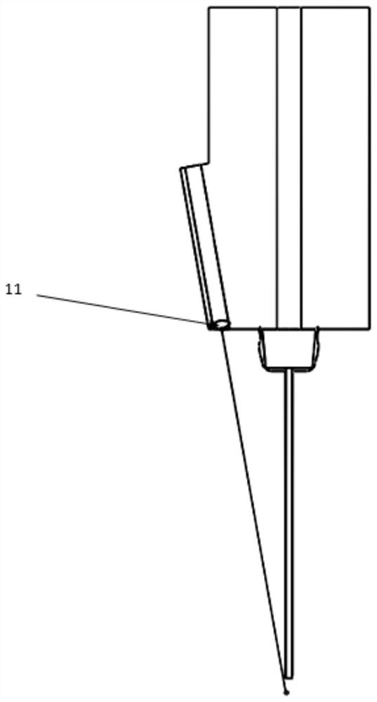
[0034] Such as figure 1 , figure 2 as well as image 3 As shown, the present invention is an in-situ 3D printing cartilage repair device, including a mechanical arm 1, an adapter frame 2, a blue light source 3, an optical fiber 4, a module 5, a pushing plate 6, a pressing plate 7, a syringe 8, and a three-way head 9. The injection needle 10 and the lens 11, the lens 11 is fixed inside the tee head 9 through a U-shaped slot, the tee head 9 is threaded to the optical fiber 4, and the optical fiber 4 is threaded to the blue light source 3 , the blue light source 3 is connected to the adapter frame 2 through bolts, the module 5 is connected to the adapter frame 2 through bolts and nuts, the push plate 6 is connected to the module 5 through bolts and nuts, and the syringe 8 is connected to the adapter frame 2 through bolts and nuts. The module is connected by a pressure plate 7, the injection needle 10 is connected to the syringe 8 by threads, the adapter frame 2 is connected to...


PUM


Abstract

The invention discloses an in-situ 3D printing cartilage repair device and method. The device comprises a lens, a raw material container, a curing light source, an adapter frame, an optical fiber, a three-way head, a pushing plate, a module, an injection needle, a pressing plate and a mechanical arm; the lens is fixedly arranged inside the three-way head through a U-shaped clamping groove, the three-way head is connected with the optical fiber through threads, the optical fiber is connected with the curing light source through threads, and the curing light source is connected with the adapter frame through bolts; the module is connected with the adapter frame through bolts and nuts, the pushing plate is connected with the module through bolts and nuts, and the adapter frame is connected with the mechanical arm through bolts; and the raw material container is connected with the module through the pressing plate, and the injection needle is connected with the raw material container through threads. According to the in-situ 3D printing cartilage repair device and method, the six-axis mechanical arm is controlled through a digital means to drive a printing spray head to directly print and repair cartilage defects in situ, the process is simple, the manual intervention time is short, and high-precision in-situ 3D printing accurate repair of various biological materials and various structures can be achieved.

Description

technical field [0001] The invention belongs to the technical field of additive manufacturing, and in particular relates to an in-situ 3D printing cartilage repair device and method. Background technique [0002] Traditional cartilage repair techniques include bone marrow stimulation and mosaic plasty. Bone marrow stimulation stimulates cartilage healing through drilling, abrasion or microfracture of the subchondral bone. Mosaic plasty fills the damaged cartilage by excavating non-moving articular cartilage to achieve Purpose of cartilage healing. Traditional cartilage repair technology is a destructive way to achieve the purpose of cartilage repair, which has certain damage to the human body. [0003] The new cartilage repair technology is a tissue engineering technology, in which biomaterials inoculated with chondrocytes are added to the damaged cartilage and implanted directly. The existing technology is mainly based on the cartilage repair method of hand-held 3D printi...

Claims


Application Information

Patent Timeline
IPC IPC(8): B29C64/135B29C64/20B29C64/209B29C64/264B29C64/321B29C64/236B29C64/393B33Y10/00B33Y30/00B33Y40/00B33Y50/02A61F2/28
Inventor 宋长辉李意浓雷浩杨余家阔杨永强
Owner SOUTH CHINA UNIV OF TECH
Learn More