Ultrasonic endoscope, artificial intelligence auxiliary identification method and system, terminal and medium

A technology of artificial intelligence and ultrasound, applied in the field of medical artificial intelligence, can solve the problems of inaccurate identification of images of stromal tumors and leiomyomas, low qualified rate of material collection, application of artificial intelligence diagnosis, etc.

Inactive Publication Date: 2021-05-14
THE AFFILIATED HOSPITAL OF QINGDAO UNIV
View PDF6 Cites 4 Cited by
  • Summary
  • Abstract
  • Description
  • Claims
  • Application Information

AI Technical Summary

Problems solved by technology

[0006] (1) Existing imaging detection or identification methods cannot accurately identify images of stromal tumors and leiomyomas, resulting in a high rate of misdiagnosis
[0007] (2) Human tissue biopsy under endoscopy is difficult to operate, especially for small lesions (diameter < 20mm), the qualified rate of specimens is low, resulting in low diagnostic rate and unclear diagnostic conclusion; at the same time, biopsy is an invasive examination, Risk of bleeding, gastrointestinal perforation
[0008] (3) At present, there is no artificial intelligence diagnosis application for endoscopic ultrasonography, especially for the identification of stromal tumors

Method used


Image

  • Ultrasonic endoscope, artificial intelligence auxiliary identification method and system, terminal and medium
  • Ultrasonic endoscope, artificial intelligence auxiliary identification method and system, terminal and medium
  • Ultrasonic endoscope, artificial intelligence auxiliary identification method and system, terminal and medium

Examples


Embodiment Construction

[0053] In order to make the object, technical solution and advantages of the present invention more clear, the present invention will be further described in detail below in conjunction with the examples. It should be understood that the specific embodiments described here are only used to explain the present invention, not to limit the present invention.

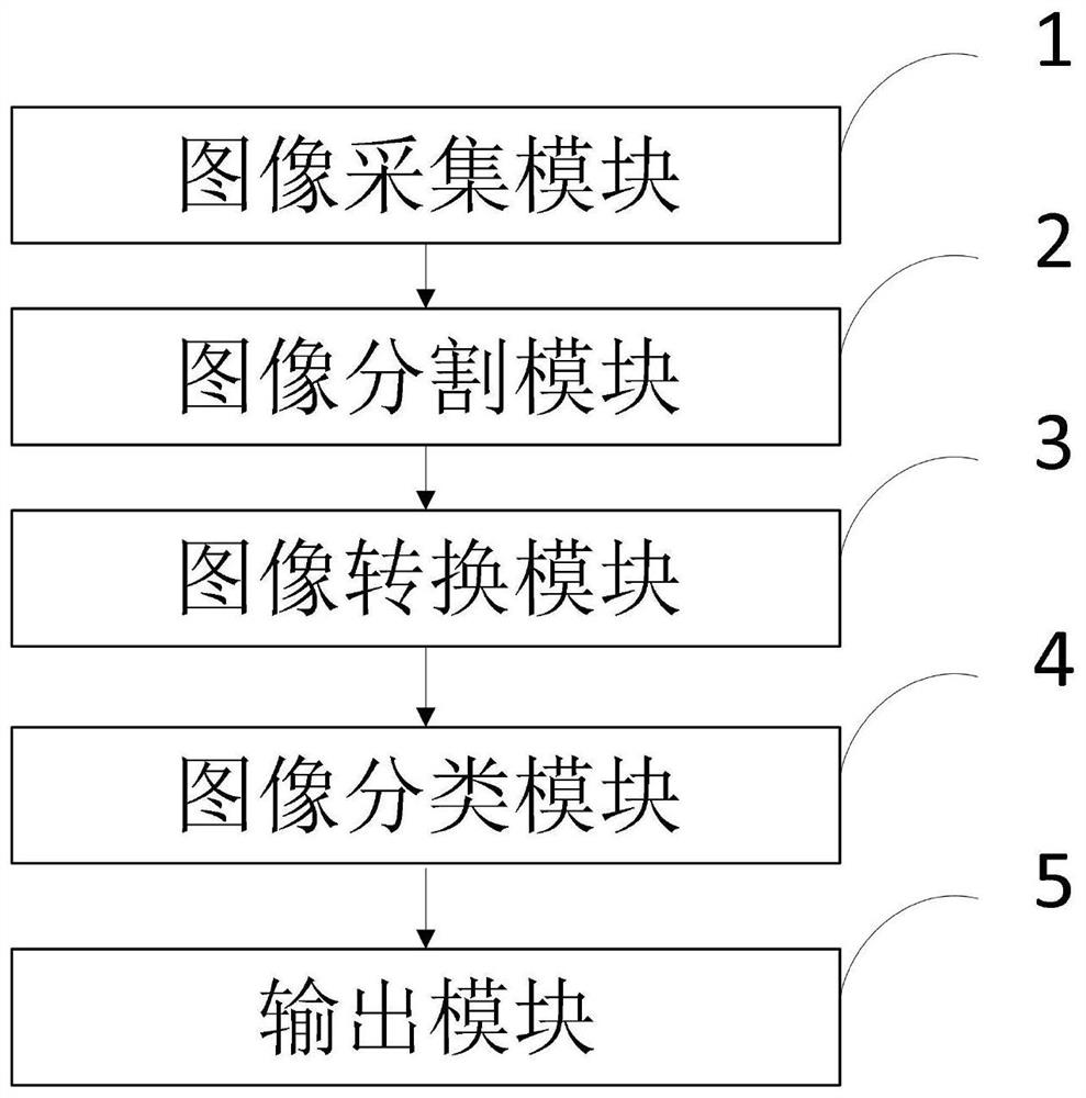
[0054] Aiming at the problems existing in the prior art, the present invention provides an ultrasonic endoscope, an artificial intelligence-assisted identification method, a system, a terminal, and a medium. The present invention will be described in detail below with reference to the accompanying drawings.

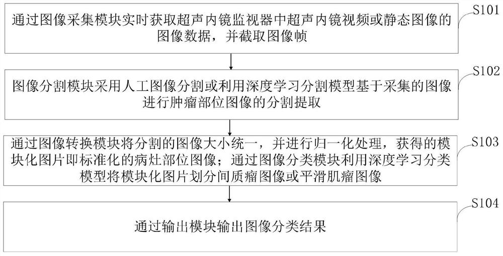
[0055] Such as figure 1 As shown, the artificial intelligence-assisted identification method under endoscopic ultrasound for stromal tumors and leiomyomas provided by the embodiments of the present invention includes the following steps:

[0056] S101, acquire the image data of the ultrasonic endoscope video or static...


PUM


Abstract

The invention belongs to the technical field of medical artificial intelligence, and discloses an ultrasonic endoscope, an artificial intelligence auxiliary identification method and system, a terminal and a medium, and the method comprises the steps: obtaining the image data of an ultrasonic endoscope video or a static image in an ultrasonic endoscope monitor in real time through an image collection module, and intercepting an image frame; segmenting and extracting the tumor part image based on the acquired image by adopting artificial image segmentation or utilizing a deep learning segmentation model through an image segmentation module; unifying the sizes of the segmented images through an image conversion module, and carrying out normalization processing to obtain a modularized picture, namely a standardized lesion part image; dividing the modular picture into an interstitial tumor image or a smooth myoma image by using a deep learning classification model through an image classification module; and outputting an image classification result through an output module. According to the method provided by the invention, the image identification accuracy can be effectively improved, and misdiagnosis is reduced.

Description

technical field [0001] The invention belongs to the technical field of medical artificial intelligence, and in particular relates to an ultrasonic endoscope, an artificial intelligence-assisted identification method, a system, a terminal, and a medium, and in particular to an artificial intelligence-assisted identification method for stromal tumors and leiomyomas under an ultrasonic endoscope . Background technique [0002] At present, endoscopic ultrasonography can clearly display the layers of the gastrointestinal tract wall and the origin of submucosal lesions. It is currently the most accurate imaging technique for diagnosing gastrointestinal submucosal tumors and is widely used in gastrointestinal submucosal lesions. Cancer screening and diagnosis. [0003] Stromal tumors and leiomyomas are the most common tumors in the submucosal tumors of the gastrointestinal tract. Stromal tumors are malignant or potentially malignant tumors that require close follow-up and surgica...

Claims


Application Information

Patent Timeline
Patent Type & Authority Applications(China)
IPC IPC(8): G06T7/00G06T7/11G06T7/62G06N3/08G16H50/20
CPCG06N3/08G06T7/0012G06T2207/10068G06T2207/10132G06T2207/20081G06T2207/20084G06T2207/30092G06T2207/30096G06T7/11G06T7/62G16H50/20
Inventor 李晓宇杨新天王晗董蒨
Owner THE AFFILIATED HOSPITAL OF QINGDAO UNIV
Why Eureka
Try Eureka