Blood vessel segmentation method, electronic device and storage medium

A blood vessel and blood vessel stenosis technology, which is applied in the field of blood vessel segmentation method, electronic device and storage medium, can solve the problem of blood vessel segmentation that cannot be refined, and achieve the effect of improving accuracy and efficiency

Pending Publication Date: 2021-05-28
SHANGHAI UNITED IMAGING INTELLIGENT MEDICAL TECH CO LTD
View PDF0 Cites 3 Cited by
  • Summary
  • Abstract
  • Description
  • Claims
  • Application Information

AI Technical Summary

Problems solved by technology

[0004] In this embodiment, a blood vessel segmentation method, an electronic device and a storage medium

Method used

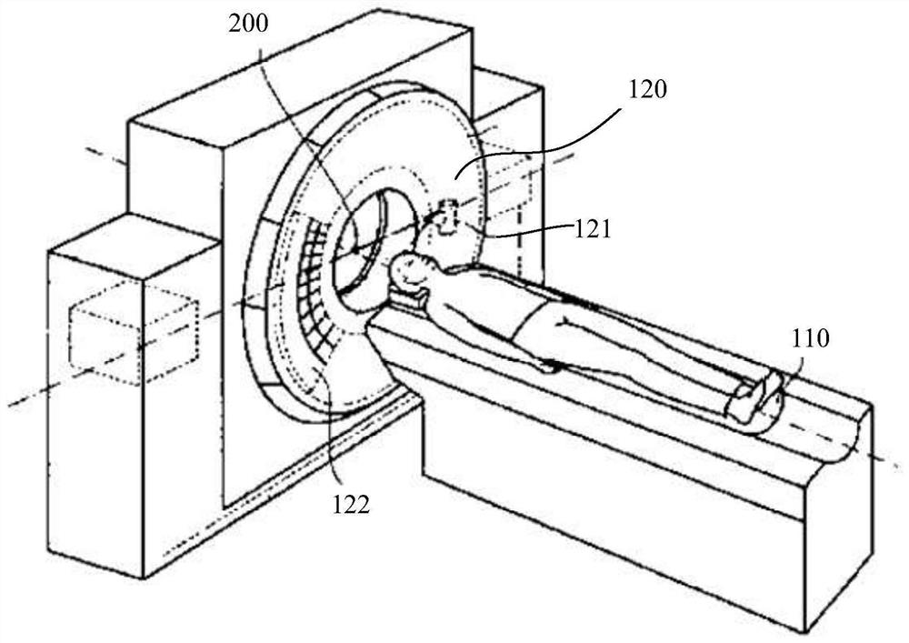
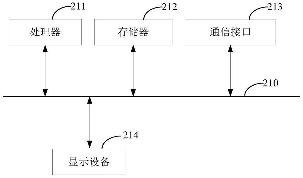

Image

  • Blood vessel segmentation method, electronic device and storage medium
  • Blood vessel segmentation method, electronic device and storage medium
  • Blood vessel segmentation method, electronic device and storage medium

Examples


Embodiment Construction

[0029] In order to understand the purpose, technical solution and advantages of the present application more clearly, the present application is described and illustrated below in conjunction with the accompanying drawings and embodiments.

[0030] Unless otherwise defined, the technical terms or scientific terms involved in the application shall have the general meanings understood by those skilled in the technical field to which the application belongs. In this application, words like "a", "an", "an", "the", "these" and the like do not denote quantitative limitations, and they may be singular or plural. The terms "comprising", "comprising", "having" and any variants thereof referred to in this application are intended to cover non-exclusive inclusion; for example, processes, methods and The system, product or device is not limited to the steps or modules (units) listed, but may include steps or modules (units) not listed, or may include other steps or modules inherent to the...


PUM


Abstract

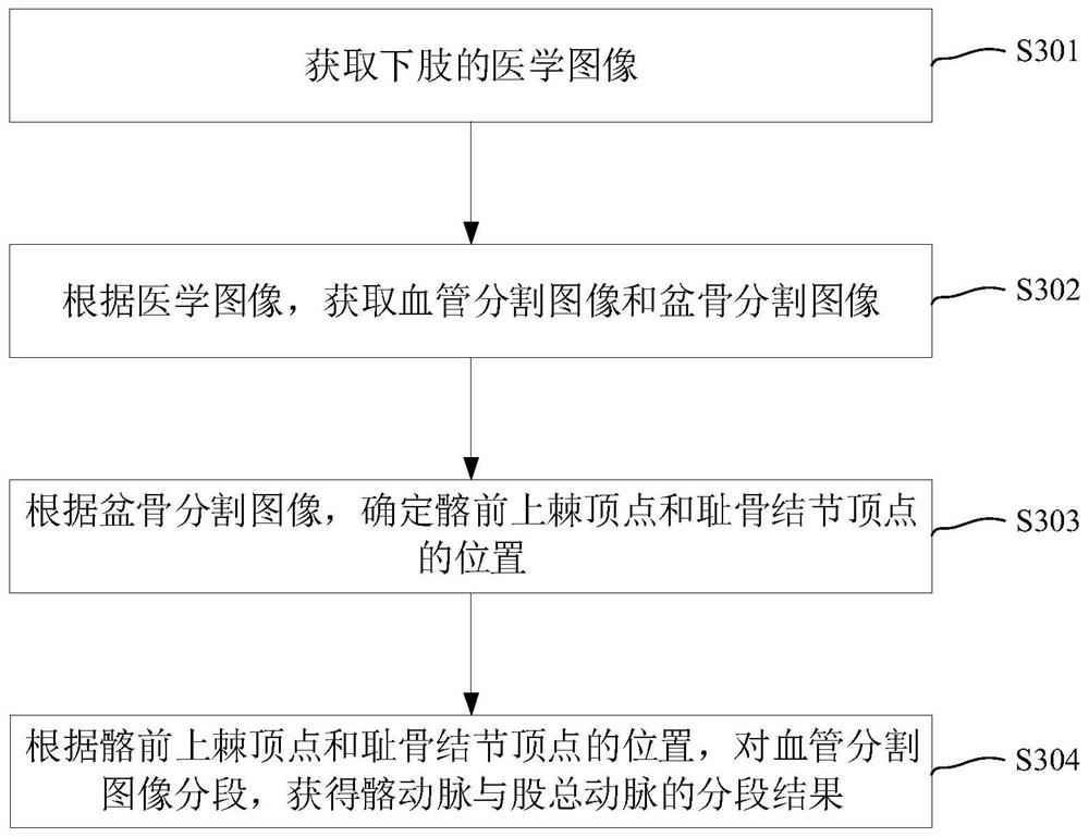
The invention relates to a blood vessel segmentation method, an electronic device and a storage medium. The blood vessel segmentation method comprises the following steps: acquiring a medical image of a lower limb; acquiring a blood vessel segmentation image and a pelvic bone segmentation image according to the medical image; determining positions of an anterior superior spine vertex and a pubic tuberosity vertex according to the pelvis segmentation image; and according to the positions of the anterior superior spine vertex and the pubic tubercle vertex, segmenting the blood vessel segmentation image to obtain segmentation results of the iliac artery and the common femoral artery. Through the blood vessel segmentation method, the defect that the iliac artery and the common femoral artery cannot be accurately classified in the prior art is overcome, the blood vessel segmentation precision is improved, full-automatic blood vessel segmentation can be achieved through a computer, and the blood vessel segmentation efficiency is improved.

Description

technical field [0001] The present application relates to the field of medical image processing, in particular to a blood vessel segmentation method, an electronic device and a storage medium. Background technique [0002] As an interventional detection method, CT angiography is widely used in the diagnosis and treatment of peripheral blood vessels and the planning of stent surgery. After computer processing of CTA images, the three-dimensional reconstructed images of peripheral blood vessels can be displayed, and the images can provide important diagnostic basis for peripheral vascular lesions. [0003] The commonly used methods of peripheral blood vessel analysis software in the current hospital system are mainly traditional image processing methods, such as threshold segmentation methods or region growing methods. However, in the process of automatic reconstruction of peripheral blood vessels, these methods will lead to abnormal conditions such as noise and fracture, and...

Claims


Application Information

Patent Timeline
IPC IPC(8): G06T7/11G06T7/00G06N3/04G06N3/08A61B6/00
CPCG06T7/11G06T7/0012G06N3/08A61B6/504A61B6/5211A61B6/5235G06T2207/10081G06T2207/10088G06T2207/10116G06T2207/10104G06T2207/10084G06T2207/30101G06T2207/20081G06T2207/20084G06N3/045
Inventor 李想沈宏
Owner SHANGHAI UNITED IMAGING INTELLIGENT MEDICAL TECH CO LTD
Why Eureka
Try Eureka