A method for isolating placental trophoblast cells from exfoliated cells of the cervix of pregnant women
A technology of trophoblast cells and exfoliated cells, which is applied in the field of separation and screening of trophoblast cells, can solve problems such as limited antibody combinations, and achieve the effects of early sampling time, good specificity, and low risk of infection and abortion
- Summary
- Abstract
- Description
- Claims
- Application Information
AI Technical Summary
Problems solved by technology
Method used
Image
Examples
Embodiment 1
[0047] Example 1 Microfluidic Sorting Chip Design
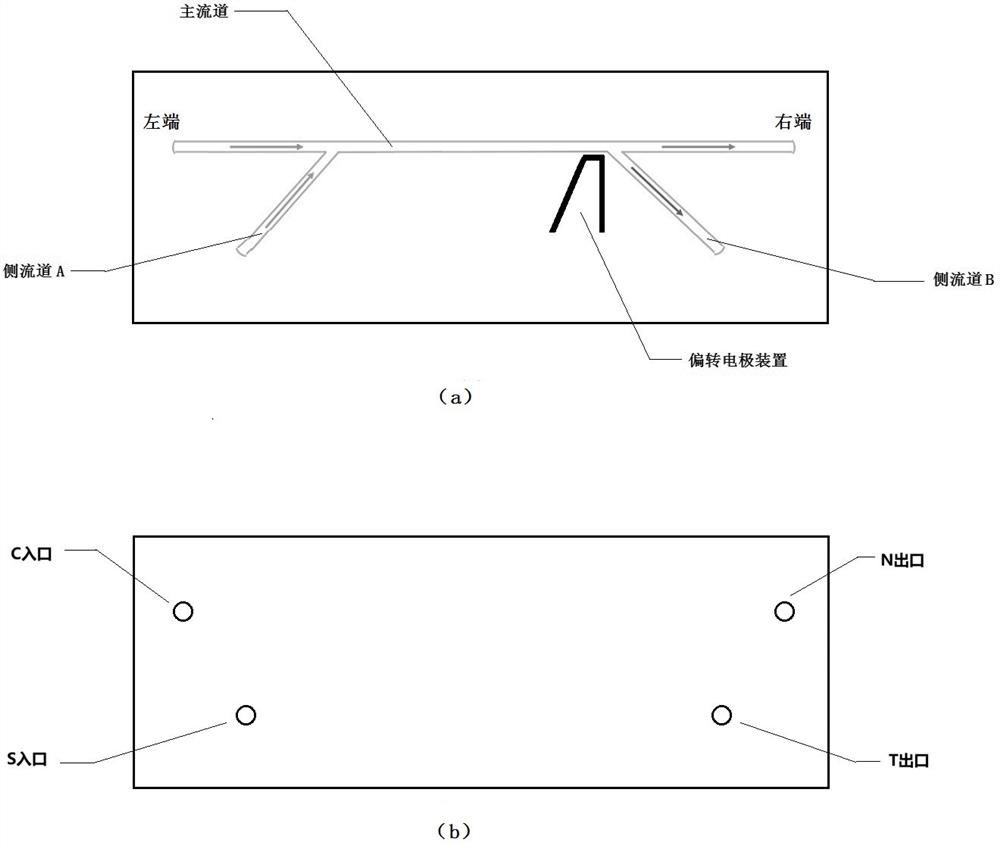
[0048] Schematic diagram of the microfluidic sorting chip used to separate and screen trophoblast cells from the exfoliated cells of the cervix of pregnant women. figure 1 shown.
[0049] The structural design of the microfluidic sorting chip is described as follows: the chip is prepared using but not limited to acrylic as the basic material, and is formed on one side of the substrate by injection molding technology. figure 1 For the shape of the pipe, the width of the pipe does not exceed 1000 μm, and the depth does not exceed 500 μm, and the other side of the substrate is used for lamination to form a complete chip.
[0050] Specifically, the microfluidic sorting chip includes a substrate and a cover attached thereto;
[0051] Such as figure 1 As shown, one side of the substrate is provided with a main channel, a side channel A and a side channel B, and the two side channels are respectively close to the left and right end...
Embodiment 2
[0057] Example 2 Separation and screening of trophoblast cells from cervical exfoliated cells of pregnant women based on microfluidic sorting chip
[0058] One, the sorting method of trophoblast cell comprises the following steps:
[0059] 1. Prepare the cervical exfoliated cell fluid sample into a sample cell suspension; the specific methods include as shown in (1)-(5) below;
[0060] 2. Add specific antibodies to the sample cell suspension for incubation; specific methods include as shown in (6)-(14) below;
[0061] 3. Using the microfluidic sorting chip of Example 1, the cell suspension incubated in step 2 was subjected to fluorescence-labeled microfluidic cell sorting; specific methods include as shown in (15)-(18) below.
[0062] Wherein, the combination of specific antibodies is shown in Table 1:
[0063] Table 1: Combinations of antigens and antibodies expressed on trophoblast cells
[0064]
[0065] Two, specifically, the sorting method of trophoblast cells compr...
Embodiment 3
[0084] Example 3 Method for Separating and Screening Trophoblast Cells from Pregnant Women's Cervical Exfoliated Cells Based on Flow Cytometry
[0085] The sorting method of trophoblast cells comprises the following steps:
[0086] 1. The cervical exfoliated cell liquid sample is prepared into a sample cell suspension; the specific method is shown in (1)-(5) in Example 2;
[0087] 2. Add specific antibodies to the sample cell suspension and incubate; the specific method is the same as shown in (6)-(14) in Example 2; wherein, the combination of specific antibodies is the same as shown in the above table 1;
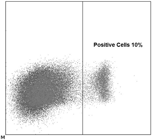
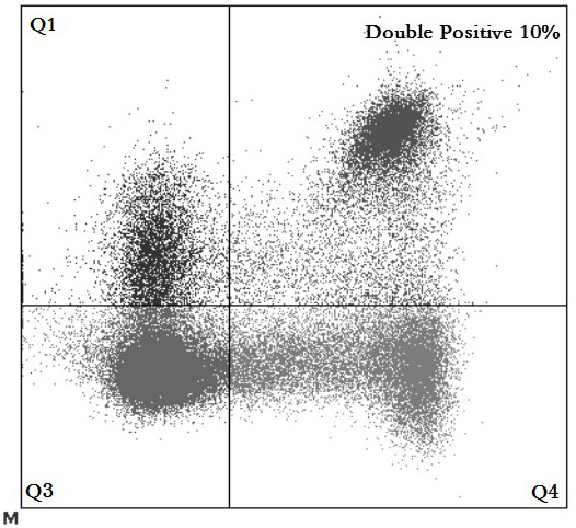
[0088] 3. Use a flow cytometry sorter (BDFACSAria II, USA) to perform fluorescent marker sorting on the cell resuspension incubated in step 2, including the following steps:
[0089] 1) Turn on the flow cytometer, and follow the operating instructions for daily start-up operations;
[0090] 2) Adjust the liquid flow of the instrument so that the breakpoint of the liquid f...
PUM
 Abstract
Description
Claims
Application Information
 - R&D
- Intellectual Property
- Life Sciences
- Materials
- Tech Scout
- Unparalleled Data Quality
- Higher Quality Content
- 60% Fewer Hallucinations
Browse by: Latest US Patents, China's latest patents, Technical Efficacy Thesaurus, Application Domain, Technology Topic, Popular Technical Reports.
© 2025 PatSnap. All rights reserved.Legal|Privacy policy|Modern Slavery Act Transparency Statement|Sitemap|About US| Contact US: help@patsnap.com


