In-vitro photoacoustic scanner

A scanning device and sound wave technology, applied in medical science, sensors, catheters, etc., can solve problems such as adverse reactions, allergic reactions, and thyroid poisoning of patients

Inactive Publication Date: 2021-07-02
康波浩瀚(北京)科技有限公司
View PDF4 Cites 0 Cited by
  • Summary
  • Abstract
  • Description
  • Claims
  • Application Information

AI Technical Summary

Problems solved by technology

However, the use of contrast media may cause adverse reactions in patients, such as thyroid poisoning or allergic reactions in patients

Method used


Image

  • In-vitro photoacoustic scanner
  • In-vitro photoacoustic scanner
  • In-vitro photoacoustic scanner

Examples


Embodiment Construction

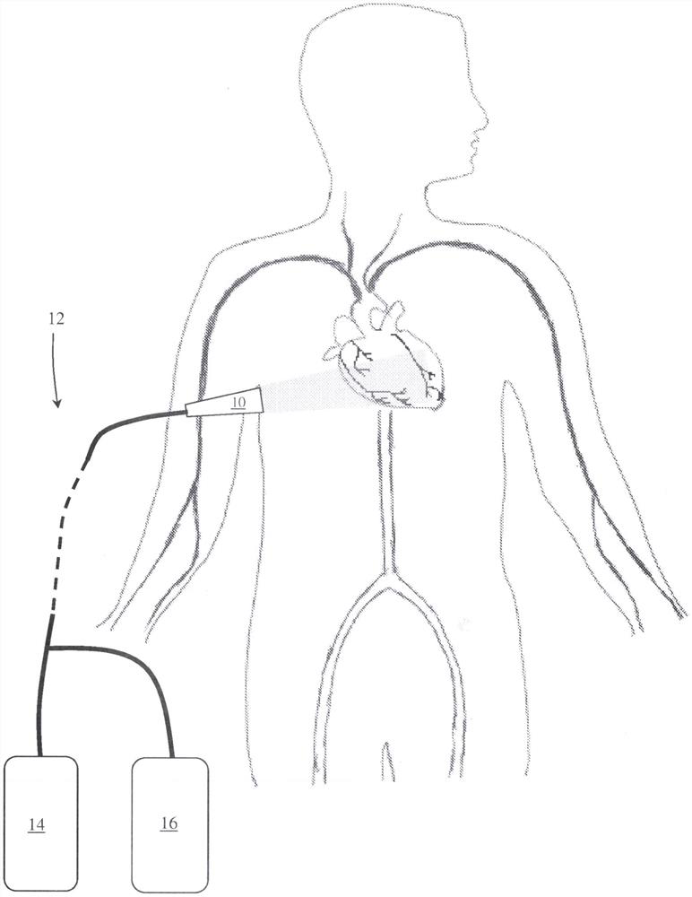
[0041] First draw attention to figure 1 middle, figure 1 A part of the human cardiovascular system is schematically shown. According to one embodiment of the present invention, a method for in vitro imaging is provided. The in vitro imaging method includes: an in vitro imaging device 10, which can be used for scanning and imaging of the cardiovascular system and internal treatment devices, figure 1 Here is the heart.

[0042] In an embodiment, the in vitro imaging device 10 may be a photoacoustic device arranged to non-invasively scan the cardiovascular system from outside the body.

[0043] The imaging device 10 in its photoacoustic embodiment may be arranged to emit laser pulses into biological tissue which, when absorbed by the biological tissue, are converted into heat generating ultrasound waves which may be detected by ultrasound waves within the device 10. The transducer detects and is analyzed to generate scan information such as: images, three-dimensional data, an...


PUM


Abstract

The invention provides an in-vitro photoacoustic scanning device which can be used for scanning and imaging human organs and body vasculature, and a treatment device. A system involved in the invention comprises an extracorporeal photoacoustic scanning device for scanning from outside, a photoacoustic device and an external display, wherein the extracorporeal photoacoustic scanning device comprises a laser and a transducer, the laser is arranged to emit laser pulses to a human organ system, the transducer is used for detecting sound waves received from the organ system so as to realize imaging of a human organ system, and a computer is used for displaying images by using special software for image processing. The device can also be used in combination with a device for opening and cleaning artery occlusion so as to achieve the purpose of in-vitro visual obstacle removal.

Description

technical field [0001] The invention relates to an extracorporeal photoacoustic scanner, which is mainly used for scanning and imaging human organs, body vascular system and internal treatment devices. Background technique [0002] Minimally invasive procedures to treat the body's vasculature or blood vessels of the body may involve delivering an interventional diagnostic or therapeutic device, such as a catheter, to a corresponding location within the body's blood vessels. The catheter is typically arranged to be entered through the body vessel or a natural orifice or stoma of a body vessel. The only minimally invasive procedure known to treat the disease in the form of a catheter is through the deployment of a stent to relieve coronary artery blockages, known as stenosis, caused by blood clots that restrict or block blood flow caused by accumulation. Commonly used therapeutic devices mainly include imaging catheters, typically those used in intravascular ultrasound (IVUS...

Claims


Application Information

Patent Timeline
Patent Type & Authority Applications(China)
IPC IPC(8): A61B5/00
CPCA61B5/0095A61B5/0084A61B5/0062
Inventor 王锋
Owner 康波浩瀚(北京)科技有限公司
Why Eureka
Try Eureka