
An anterior segment three-dimensional ultrasound scanning imaging device and method

A technology of scanning imaging and three-dimensional ultrasound, which is applied in the directions of ultrasound/sonic/infrasonic image/data processing, ultrasound/sonic/infrasonic diagnosis, eye examination, etc., and can solve image resolution degradation, unfavorable room angle accurate measurement, and information loss and other problems, to achieve the effect of improving the reconstruction image quality and measurement accuracy, alleviating the degree of geometric distortion, and avoiding artifact interference

Active Publication Date: 2021-12-07
INST OF BIOMEDICAL ENG CHINESE ACAD OF MEDICAL SCI
View PDF1 Cites 0 Cited by
  • Summary
  • Abstract
  • Description
  • Claims
  • Application Information

AI Technical Summary

Problems solved by technology

However, because the structure is too complex and expensive, it is difficult to promote the use in clinical ophthalmology
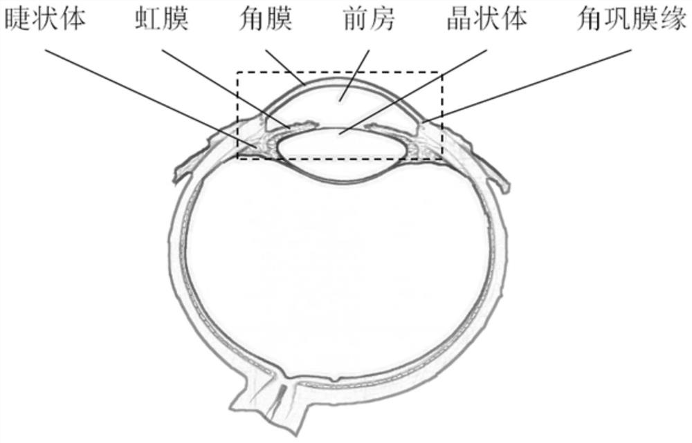
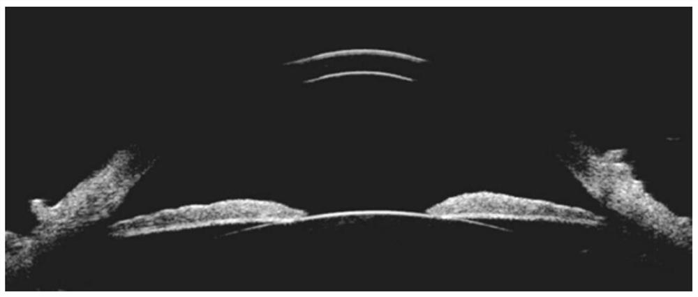
[0005] The structure of linear scanning is relatively complex, but the resolution difference and geometric distortion of the obtained anterior segment ultrasound images will be improved to a certain extent. It is currently the main scanning method clinically used in anterior segment ultrasound diagnosis. The commonly used methods include horizontal linear panorama There are two types of scanning and lateral linear scanning. The advantage of horizontal linear scanning is that it can display the section image of the anterior segment tissue in a panoramic view in one image, observe the iris and chamber angle on both sides at the same time, and measure the central corneal thickness, pupil diameter, and anterior chamber depth. Please refer to figure 2 , figure 2 It is a schematic diagram of horizontal linear panoramic scanning imaging. Since the front and rear surfaces of the cornea are smooth tissue interfaces, it is difficult for the sound wave to be received by the transducer after being reflected near the corneoscleral limbus. figure 2 Artifacts shown in ; at the same time, this scanning method is also not conducive to the accurate measurement of the chamber angle and the display of deeper tissues such as the ciliary body
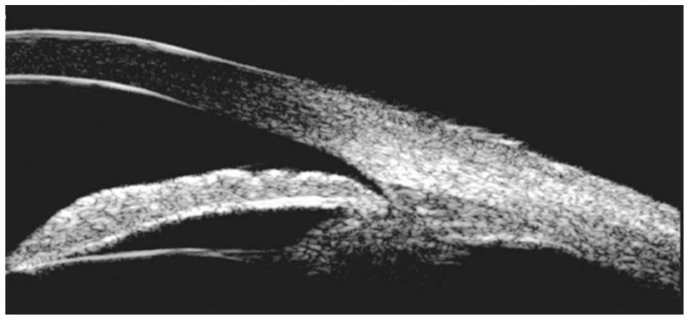
Lateral linear scanning can more clearly display the front and rear interfaces of the iris, ciliary body and other structures, and can accurately measure the opening and closing of the room angle, please refer to image 3 , image 3 It is a schematic diagram of lateral linear scan imaging. As shown in the figure, lateral linear scan imaging cannot observe the entire anterior segment, and cannot complete biometric measurements in the direction of the eye axis
[0006] Also in the prior art, the reciprocating motion of the stepping motor is used to scan the two-dimensional image, and the two-dimensional image is used to construct a three-dimensional image (such as CN103815931A), but the imaging rate of this mechanical scanning method is low, and a three-dimensional image is completed once. Reconstruction takes a long time, and it requires the patient to keep the eye as still as possible during the scanning process, so the patient needs to have a strong ability to cooperate, and it puts forward higher requirements for later image registration, segmentation, reconstruction, etc. , Moreover, in order to ensure the real-time performance of 3D imaging, it is necessary to reduce the amount of 2D image collection, resulting in insufficient raw data, missing information, and reduced image resolution

Method used


Image

  • An anterior segment three-dimensional ultrasound scanning imaging device and method
  • An anterior segment three-dimensional ultrasound scanning imaging device and method
  • An anterior segment three-dimensional ultrasound scanning imaging device and method

Examples


Embodiment 1

[0039] Please refer to Figure 4 , Figure 4 It is a structural exploded diagram of the three-dimensional ultrasonic scanning imaging device of the anterior segment described in Embodiment 1 of the present invention. Embodiment 1 of the present invention provides a three-dimensional ultrasonic scanning imaging device for the anterior segment, which includes a three-dimensional ultrasonic scanning imaging probe and a high-frequency linear array transducer (not shown). The three-dimensional ultrasonic scanning imaging probe includes a gear assembly, a probe base 20, a transducer base 30 and a driving assembly.

[0040] The gear assembly includes a gear seat 11 , a spur gear 12 , a sector gear 13 and a positioning pin 14 . The gear seat 11 is a hollow structure. Specifically, in this embodiment, it includes a top plate 111 and a first side wall 112 and a second side wall 113 vertically fixed on both sides of the top plate 111; the spur gear 12 and the sector gear 13 Mesh with ...

Embodiment 2

[0049] Embodiment 2 of the present invention provides a method for three-dimensional ultrasonic scanning imaging of the anterior segment, which is performed using the three-dimensional ultrasonic scanning imaging device for the anterior segment described in Embodiment 1, and specifically includes the following specific steps:

[0050] S1. Make the first rotation axis correspond to the eye axis of the scanned eyeball, the second drive motor 50 drives the spur gear 12 to rotate around the second rotation axis, the spur gear 12 meshes to drive the sector gear 13 to rotate, and makes the The high-frequency linear array transducer forms an included angle with the eye axis of the scanned eye, and the included angle is 0-30°;

[0051] S2. The first driving motor 40 drives the gear seat 11 to rotate around the first rotation axis for one rotation, and makes the high-frequency linear array transducer rotate one rotation along the tilt angle in front of the scanned eyeball to form a coni...


PUM


Abstract

The present invention relates to a three-dimensional ultrasonic scanning imaging device and method for the anterior segment. The three-dimensional ultrasonic scanning imaging device for the anterior segment includes a three-dimensional ultrasonic scanning imaging probe, which includes a gear assembly, a probe seat, a transducer seat and a driving assembly; the gear assembly includes gear seat, spur gear and sector gear; the drive assembly includes a first drive motor and a second drive motor, the second drive drive motor is fixedly connected to the spur gear, and can drive the spur gear to rotate around the second rotation axis and drive the sector The gears are deflected, so that the transducer seat and the high-frequency linear array transducer form an inclined angle, and the first drive motor is used to drive the gear assembly and other components to rotate, so that the high-frequency linear array transducer is in front of the eyeball to be scanned. The side rotates one circle along the tilt angle to form a conical plane, so that various tissue structures can be better visualized, artifact interference can be avoided, and biological measurements such as the length, angle, area, and volume of each structural position can be completed. .

Description

technical field [0001] The invention relates to the technical field of laser ultrasonic nondestructive testing, in particular to a three-dimensional ultrasonic scanning imaging device and method of the anterior segment. Background technique [0002] In the concept of medical eye anatomy, the anterior segment includes the cornea, anterior chamber and lens, and is the main component of the refractive system of the eyeball. In recent years, the prevalence of the anterior segment has been increasing year by year, so the examination of the cornea is particularly important. At the same time, the condition of the lens is also an important reference in the diagnosis of glaucoma, cataract and refractive vision problems. Due to the superficial position of the eyeball and orbit, regular structure, clear interface, and less sound attenuation, it is suitable to use ultrasound for inspection and diagnosis, and because it does not require high penetration and high resolution requirements, ...

Claims


Application Information

Patent Timeline
Patent Type & Authority Patents(China)
IPC IPC(8): A61B8/10
CPCA61B8/10A61B8/466A61B8/52A61B8/5223
Inventor 王晓春周盛杨军计建军王文赛巩丽文
Owner INST OF BIOMEDICAL ENG CHINESE ACAD OF MEDICAL SCI
Learn More