Abnormal degree quantification method of gastric mucosa dyeing magnified image microstructure

A technology of magnifying images and quantifying methods, applied in the field of image processing in the medical field, can solve problems such as learning difficulties, and achieve the effect of improving reliability and accuracy

Active Publication Date: 2021-09-03
WUHAN UNIV
View PDF3 Cites 5 Cited by
  • Summary
  • Abstract
  • Description
  • Claims
  • Application Information

AI Technical Summary

Problems solved by technology

However, whether the shape of the microstructure is uniform, whether the distribution is symmetrical, etc., are all qualitative descriptions or experience summaries based on the experience of early cancer diagnosis by endoscopists. Quantitative calculation methods and corresponding conclusions are not given for each description, which has great significance. Strong subjectivity, learning difficulties

Method used


Image

  • Abnormal degree quantification method of gastric mucosa dyeing magnified image microstructure
  • Abnormal degree quantification method of gastric mucosa dyeing magnified image microstructure
  • Abnormal degree quantification method of gastric mucosa dyeing magnified image microstructure

Examples


Embodiment Construction

[0051] The technical solution of the present invention will be further described in detail below in conjunction with the accompanying drawings and specific embodiments.

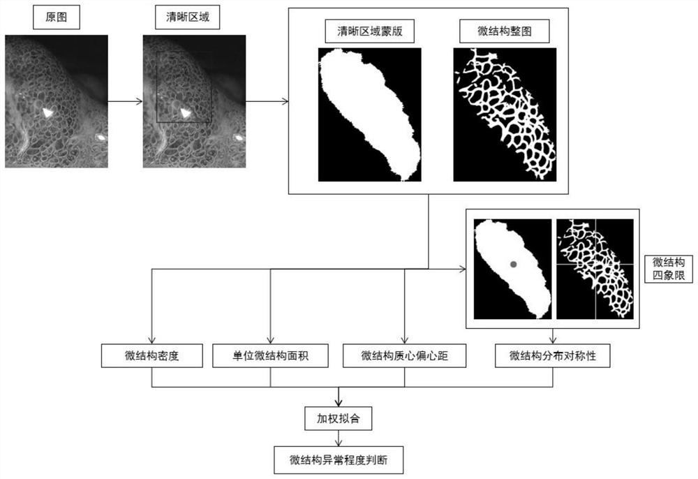
[0052] Please refer to figure 1 , a schematic diagram of the implementation of the method for quantifying the abnormality degree of the microstructure of the enlarged image of gastric mucosa staining proposed by the present invention. Such as figure 1 As shown, the method for quantifying the abnormality degree of the microstructure of the enlarged image of gastric mucosa staining of the present invention includes a calculation method of microstructure density, a calculation method of unit microstructure area, a calculation method of microstructure centroid eccentricity, and a calculation method of microstructure distribution symmetry.

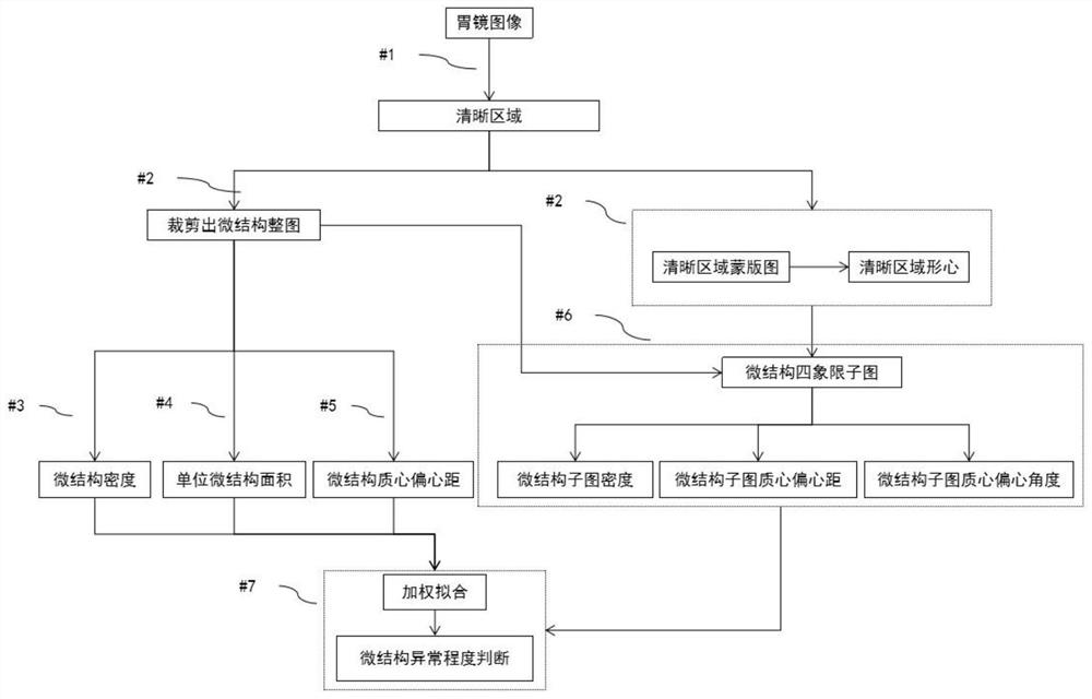
[0053] Please refer to figure 2 , which takes the embodiment of the method for quantifying the abnormality degree of the microstructure of the enlarged image of gastric muc...


PUM


Abstract

The invention discloses an abnormal degree quantification method of gastric mucosa dyeing magnified image microstructure. The method comprises an image segmentation method, a density method, a unit area method, a centroid eccentricity method and a distribution symmetry method. The image segmentation method is used for extracting a clear area and a microstructure whole image in the gastroscope image; the density method is used for calculating the microstructure whole graph density; the unit area method is used for calculating the area occupied by each microstructure; the centroid eccentricity method is used for calculating the offset distance of the equivalent centroid of the microstructure whole graph relative to the centroid of the clear area; the distribution symmetry method is used for quantifying the four-quadrant symmetry of the microstructure. And finally, the microstructure density, the microstructure unit area, the microstructure centroid eccentricity and the microstructure distribution symmetry are weighted to obtain a microstructure anomaly degree coefficient, and thus judging the microstructure anomaly degree grade according to the microstructure anomaly degree coefficient.

Description

technical field [0001] The invention relates to the technical field of image processing in the medical field, in particular to a method for quantifying the abnormality degree of the microstructure of the enlarged image of gastric mucosa dyeing. Background technique [0002] Gastric cancer is one of the most common digestive tract malignancies and the third deadliest malignancy in the world. In 2015, the number of gastric cancer cases in my country reached 400,000, and the death toll was close to 300,000, seriously endangering people's lives and health. The root cause of gastric cancer endangering human health is that it cannot be detected early. The advent of chromoendoscopy established the endoscopic diagnosis method for early gastric cancer. The combined application of magnifying gastroscopy and electronic chromoendoscopy can observe the tiny blood vessel structure and the fine structure of mucosal surface that cannot be observed by ordinary gastroscopy, which provides c...

Claims


Application Information

Patent Timeline
Patent Type & Authority Applications(China)
IPC IPC(8): G06T7/00G06T7/11G06T7/62G06T7/66G06T7/68G06T11/40G06N3/04
CPCG06T7/0012G06T7/11G06T7/62G06T7/66G06T7/68G06T11/40G06T2207/30092G06T2207/30096G06T2207/30101G06N3/045
Inventor 于红刚吴练练董泽华
Owner WUHAN UNIV
Why Eureka
Try Eureka