Colloidal gold test strip capable of detecting combined compound of Tg and anti-Tg antibody

A colloidal gold test paper, colloidal gold technology, applied in the direction of biological testing, measuring devices, material inspection products, etc., can solve the problem of undetectable serum Tg content, etc.

Pending Publication Date: 2021-09-07
SUZHOU DONGNI BIOTECH CO LTD
0 Cites 2 Cited by

AI-Extracted Technical Summary

Problems solved by technology

Therefore, when DTC recoverers relapse into DTC, although the secretion of Tg increases, due to the presence of TgAb in the body, Tg...

Method used

The structure of Tg test paper strip is as shown in Figure 3 and 4, comprises rectangular bottom plate 1, and is arranged on described bottom plate 1 lapped sample pad 2, colloidal gold binding pad 3, nitrocellulose membrane 4 and Absorbent pad5. In a preferred embodiment, the interface between the sample pad 2 and the colloidal gold bonding pad 3 is enlarged to improve the diffusion efficiency of the sample. Preferably, the junction of the sample pad 2 and the colloidal gol...

Abstract

The invention relates to a colloidal gold test strip capable of detecting a combined compound of Tg and an anti-Tg antibody. The colloidal gold test strip is provided with a detection line, the colloidal gold combined pad contains colloidal gold particles labeled by a combined antibody; a capture antibody is anchored in the detection line, the binding antibody is an anti-Tg polyclonal antibody, and the capture antibody is a mouse anti-human IgG antibody. According to the invention, Tg is used for immunizing an animal which is far away from human genetic relationship, unique antiserum is obtained, and the antiserum and a mouse anti-human IgG antibody can be combined to prepare a colloidal gold test strip for detecting a Tg-anti-Tg antibody compound. By using a specific antiserum combination, the problem that a test strip prepared from single antiserum is poor in correlation in a quantitative detection process is solved. The colloidal gold test strip also has a function of detecting whole blood, and by using the colloidal gold test strip, the Tg-anti-Tg antibody compound and content in peripheral blood of a subject can be rapidly detected, and a convenient tool is provided for monitoring recurrence of a differentiated thyroid cancer convalescent patient.

Application Domain

Biological testing

Technology Topic

Polyclonal antibodiesAntigen binding +14

Image

  • Colloidal gold test strip capable of detecting combined compound of Tg and anti-Tg antibody
  • Colloidal gold test strip capable of detecting combined compound of Tg and anti-Tg antibody
  • Colloidal gold test strip capable of detecting combined compound of Tg and anti-Tg antibody

Examples

  • Experimental program(1)

Example Embodiment

[0022] The principles and features of the present invention will be described below with reference to the accompanying drawings, and the exemplary examples are intended to be construed as they are intended to limit the scope of the invention.
[0023] 1, and prepared for antibody screening
[0024] In order to study the detection antibody Tg- anti-Tg antibody complex, we tried a variety of ways.
[0025] First, try using a variety of anti-Tg monoclonal antibody, respectively Tg- anti-Tg antibody affinity complex and experimental results show that the use of all commercially available anti-Tg monoclonal antibodies and our own research and development of anti-Tg monoclonal antibody did not demonstrate effective antibody titers.
[0026] After our in-depth study of the causes, the results show that, due Tg itself caused antiserum containing anti-Tg polyclonal antibodies, these antibodies Tg complex formation, covered the vast majority of epitopes on Tg, resulting in Tg monoclonal antibody basic It is unlikely to bind to the complex. Therefore, we need to find new specific antibodies.
[0027] In order to find new specific antibodies against Tg, we used the full-length human protein Tg immune to many different species of animals, and to evaluate the anti-serum titer by using Tg- anti-Tg antibody complex.
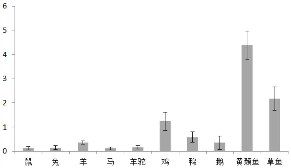
[0028] Such as figure 1 Shown, diluted 2560-fold, catfish and carp antisera has higher binding values. Compared with fish, poultry antisera binding it has a relatively low value, chicken antiserum binding value higher in poultry. Binding mammalian antisera very low values, wherein relatively high goat antiserum. In general, fish and avian antiserum antisera having a certain potency Tg- be used to detect anti-Tg antibody complex. Mammal, sheep antiserum showed some positive reaction, may be used to detect anti-Tg antibody complex Tg-.
[0029] 2, the test strip was prepared
[0030] Based on these experiments, we are ready to prepare colloidal gold strip using the method of double-antibody sandwich. To fish, birds and other anti-serum as the binding of antibodies to colloidal gold. Since the anti-Tg antibody Tg- complex anti-Tg antibody is a human antibody, therefore, select the mouse anti-human IgG antibody as the capture antibody.
[0031] The Tg of the test strip structures image 3 with 4 , Includes a rectangular base plate, said base plate and disposed in overlapping sequentially on a sample pad 2, 3 gold conjugate pad, a nitrocellulose membrane 4 and pad 5. In a preferred embodiment, the sample pad 2 bound to colloidal gold interface between the pad 3 is increased to improve the efficiency of diffusion of the sample. Preferably, the sample pad 2 bound to colloidal gold pad at the junction of the upper portion 3 made of a shape protruding, colloidal gold conjugate pad with the sample pad 3 at the junction of the lower portion 2 is made protruding shape, and the two parts fit each other, to the contact surface is increased, and, since the junction 2 on the sample pad, the colloidal gold conjugate pad 3 below, under the force of gravity facilitates the diffusion of the sample from the sample pad 2 gold conjugate pad 3.
[0032] 2 is a sample pad through the phosphate buffer treated glass fiber membranes, the phosphate buffer was phosphate buffer containing 1-5% BSA and surfactant.
[0033] 3 is a colloidal gold conjugate pad coated glass fiber membrane bound antibody colloidal gold particles. Gold conjugate pad contains gold particles 3, the colloidal gold particles bound anti-Tg polyclonal antibodies (antisera catfish, carp antiserum, antiserum chicken, duck or goose antisera antiserum). Was added dropwise in 2 sample pad after a sample if Tg- anti-Tg antibody complex, the complex diffuses into the gold conjugate pad 3, by colloidal gold particles may be present. Composite with colloidal gold particles continue to diffuse into the nitrocellulose membrane 4, and collected on nitrocellulose membranes displayed 4.
[0034] Colloidal gold particles bound labeled antibodies as follows:
[0035] 1) Preparation of colloidal gold: 1% chloroauric acid solution was diluted with distilled water to 0.01%, boil, a solution of trisodium citrate was added, continue to boil, to know a bright red liquid, heating was stopped, to make up the loss due to boiling water, to obtain colloidal gold;
[0036] 2) After the addition of colloidal gold antiserum mix was allowed to stand, the precipitate was centrifuged, washed twice, to obtain a labeled colloidal gold particles and AP8. The antiserum labeled colloidal gold particles sprayed in the glass fiber membrane, prepared as gold conjugate pad 3.
[0037] In a preferred embodiment, such as Figure 4 , The sample pad hemofilter membrane 6 is provided with the interface of the 2 3 binding to colloidal gold pad. The test strip is provided so that the present invention may be used in whole blood samples, not limited to serum or tissue eluate.
[0038] In a preferred embodiment, the hemofilter membrane labeled antibody against an erythrocyte (RBC), red blood cells can be trapped, and other substances in the blood driven electrostatic attractive force, the smooth filtration of blood through the membrane to move colloidal gold pad. Rapid diagnostic tests done with whole blood samples, which is difficult to ensure a target substance can be affected by the sample pad and the blood cells can be isolated completely out, so that the final detection result, color interference from blood cells, peripheral blood in order to achieve direct for detecting, serum was extracted from peripheral blood without the need for re-testing.
[0039] 4 is provided on the nitrocellulose membrane 411 for detecting the line. When the detection line 411 on the anchor coated with mouse anti-human IgG antibody, while the composite with colloidal gold particles is spread on a nitrocellulose membrane 4, the line 411 encounters the detection, the detection line 411 in combination with the anchor capture antibody gathered on the detector line 411, the composite with colloidal gold particles aggregate more lines in the detection, the deeper the color.
[0040]A quality control line 42 is also provided on the nitric acid fiber membrane 4, and the heat line 42 is detached from the colloidal gold bond pad 3 than the detection line 41. The secondary antibody of the heat line 412 is anchored (e.g., mouse anti-yellow squid IgM, mouse anti-grass IgM, mouse anti-chicken IgG, mouse anti-duck IgG or mouse anti-goose IgG). When the colloidal gold particles are diffused to the heat line 42, it is agreed on the heat line 42. Therefore, the colloidal gold particles are in the diffusion process, in case of the detection line 41, the colloidal gold particles combined with the TG-anti-TG antibody complex are aggregated on the detection line 41, and the colloidal gold particles without the complex continue to diffuse forward. When the quality control line 42 is encountered, it is aggregated to the quality control line 42. Since the colloidal gold particles are much greater than the complex, whether or not the Tg-anti-Tg antibody complex is present in the sample, the heat colloidal line 42 will be developed.
[0041] The preparation method of nitric acid fiber membrane 4 is as follows:
[0042] 1) Close the nitric acid fiber membrane in the blocking liquid, the blocking liquid is a phosphate buffer containing 1% BSA and 0.1% Tween-20 (pH 7.0);
[0043] 2) The capture antibody and the heat control antibody are added to the point film dilution, resulting in a capture antibody point film liquid and a heat control antibody point membrane, and the captured antibody dot film liquid and the heat gas-controlled antibody nodes are 2 μl / cm, respectively. The amount is sprayed on a 5 mm detection line and a quality control line. Among them, the dilution of the dilution is 0.01 m phosphate buffer (pH 7.4) containing 0.15 M sodium chloride, 10 mM ethylenediamine tetracetic acid, 1 g / L sodium and 25 g / L methanol (pH 7.4). The concentration of the captured antibody dot film was 3.5 μg / ml, the concentration of the heat gas-controlled antibody dot film solution was 1.5 μg / L.
[0044] When qualitative detection, 50 μl of peripheral blood, serum or tissue eluent sample was added to 50 μl of sample pretreatment solution, stir mixed, dripped in the sample pad of the sample pad of the test strip, from 20-30 ° C Lower chromatography 5 min. By observing the detection line and the quality control line, it is determined whether or not the composite is contained in the sample. That is, when the detection line and the quality control line are color, the sample contains a complex; when the detection line does not color, the quality line is color, and the sample does not contain Tg.
[0045] In a specific embodiment, the sample pretreatment liquid is a phosphate buffer containing 0.3 0.5% ammonium chloride, 0.1% potassium carbonate. This sample pretreatment liquid can crack the red blood cells in whole blood samples, which is advantageous for detection of whole blood samples.
[0046] When quantitative detection, you need to prepare the color value reading system. The test strip after the chromatography was placed under the scanning device of the color value-concentration system, and the scanned image was processed by the color value reading system to obtain the detected value, according to the corresponding relationship between the detection value and the standard curve, The concentration of the complex.
[0047] During the actual use, since the TG-anti-TG antibody complex is shorter in the peripheral blood of the subject, in order to improve the accuracy, the outer peripheral blood can be detected in 20 minutes during sampling, Prevent the window period that misses the existence of the complex in the peripheral blood.
[0048] 3, qualitative test strip results
[0049] We use the above test strip to qualitatively using the above test strips after chemical antisera (excess) containing Tg-induced antisera.
[0050] The test paper is detected using the quantitative detection test above. The results are shown in Table 1, and the test strip detection of the anti-precursor prepared by Huangjifish is less than 50 ng / ml, higher than 30 ng / ml. The test strip detection by the anti-preparation of grassfish is less than 100 ng / ml, higher than 50 ng / ml. Chicken anti-serum prepared test strip detection is less than 500 ng / ml, higher than 250 ng / ml. Duck and goose anti-preparation prepared test strip detection are less than 250 ng / ml, higher than 100 ng / ml. Since the normal value of the serum Tg in the human body is 3-40 ng / ml, the test strip with anti-preparation prepared by yellow squid and the test strip prepared by grassfish antisexum were qualitatively detected.
[0051] Table 1 Detection of test strips using different binding antibodies contains different concentrations of TG-anti-TG antibody complexes
[0052]
[0053] 4, quantitative test strip optimization
[0054] During the quantification test paper strip experiment, we found a new problem, using the anti-blood-prepared test strip of the yellow squid or the squid, there is no good linear relationship in quantitative detection. Test paper strips using the sample concentrations of 100, 125, 150, 175, 200, 225, 250 ng / ml. The test strip is then used for the color value-concentration system for standard curve establishment. Such as Figure 5 with 6 As shown, whether it is using yellow squid anti-preparation or a test strip, a test strip, related coefficient R 2 Unqualified as a concentration measurement as a concentration measurement.
[0055] Not subject to the existing theory, we speculate why, although human serum contains antibodies covering the vast epitope of TG protein, each TG protein is incubated after being incubated by serum, with a combination of the antibody therein Randomness, resulting in an epitope exposed by each Tg-anti-TG antibody complex. Some composites may expose yellow squid antisera or grassfish antisera-combined epitope, and some may not expose, thus limiting linear correlation.
[0056] To this end, we experimented a variety of antiserum combinations for preparing a test strip. It is found that an antiprapent combination of a relatively affiliated property (e.g., the antiseroff carp antiseroff, anti-precipitation of yellow squid can not make up for defects of correlation. After experimenting with a variety of combinations, we found that when the anti-precision of the yellow squid, the duck anti-serum and sheep anti-serum is combined with a striped strip prepared as a binding antibody. Such as Figure 7 As shown, a linear correlation coefficient of greater than 0.99 can be obtained using this combination, which greatly improves the correlation of the detection value and the actual value, which can be used as a quantitative test strip.
[0057] The preferred embodiments of the present invention are not intended to limit the invention, and any modifications, equivalents, improvements, and the like, which may be contained in the present invention. Within the range.

PUM

no PUM

Description & Claims & Application Information

We can also present the details of the Description, Claims and Application information to help users get a comprehensive understanding of the technical details of the patent, such as background art, summary of invention, brief description of drawings, description of embodiments, and other original content. On the other hand, users can also determine the specific scope of protection of the technology through the list of claims; as well as understand the changes in the life cycle of the technology with the presentation of the patent timeline. Login to view more.
Why Eureka
Try Eureka