Wearable visual ultrasonic non-invasive monitoring equipment

A monitoring equipment, ultrasonic technology, applied in the direction of ultrasonic/sonic/infrasonic diagnosis, ultrasonic/sonic/infrasonic Permian technology, ultrasonic/sonic/infrasonic image/data processing, etc., can solve the lack of artificial intelligence and the impact of imaging quality , Imaging and detection inconvenience and other problems, to improve the detection accuracy, the patient's health status is intuitive and effective, and the effect of reducing energy loss

Pending Publication Date: 2021-09-28
SHANGHAI SIXTH PEOPLES HOSPITAL
View PDF0 Cites 2 Cited by
  • Summary
  • Abstract
  • Description
  • Claims
  • Application Information

AI Technical Summary

Problems solved by technology

Even so, due to the rigidity of the ultrasound probe, it is often impossible to obtain good contact, which greatly affects the imaging quality
In the detection of human tissue, if it is not a soft tissue part, or when the surface curvature of the detection target changes continuously and irregularly, the probe cannot be used, which causes great inconvenience to imaging and detection. It needs to be held by the medical staff during use, so it cannot be used continuously. surveillance imaging
Second, due to the lack of artificial intelligence, automatic calculation and real-time monitoring of venous vessel area cannot be achieved

Method used


Image

  • Wearable visual ultrasonic non-invasive monitoring equipment
  • Wearable visual ultrasonic non-invasive monitoring equipment
  • Wearable visual ultrasonic non-invasive monitoring equipment

Examples


Embodiment Construction

[0023] In order to enable those skilled in the art to better understand the technical solution of the present invention, the following examples describe the present invention in further detail, and the following examples are only used to illustrate the invention, but are not intended to limit the scope of the present invention.

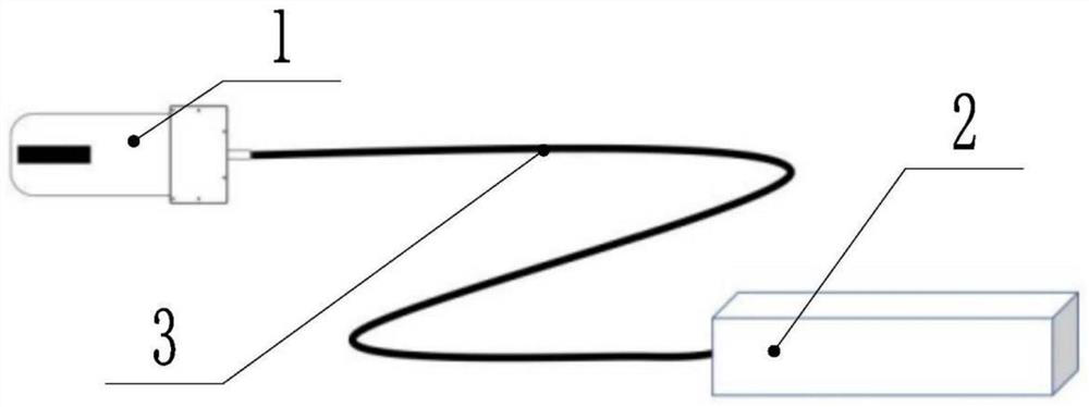
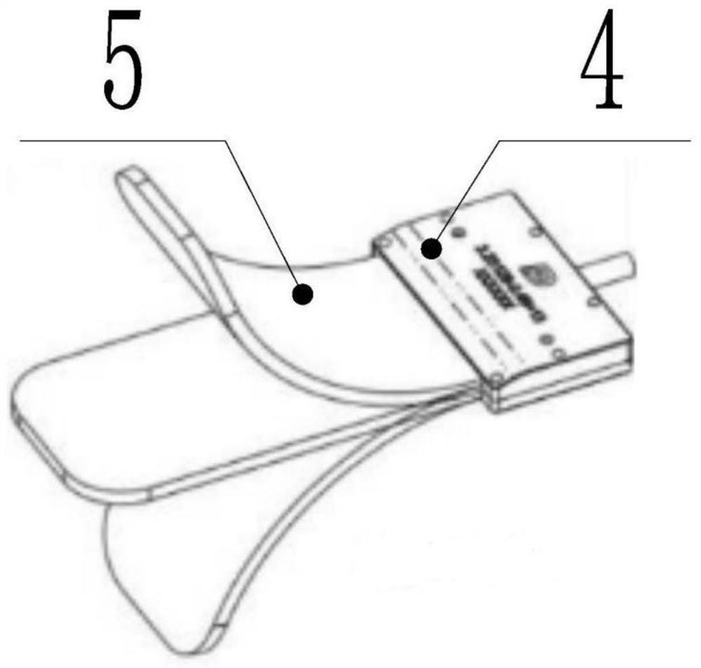
[0024] A wearable and visualized ultrasonic non-invasive monitoring device, comprising: a flexible ultrasonic probe 1, an ultrasonic scanning host 2 connected to the flexible ultrasonic probe through a connecting line 3, and the ultrasonic scanning host 2 is connected to a display device. The flexible ultrasonic probe 1 includes a flexible layer medium, and the ultrasonic transducer units are arranged in or on the surface of the flexible medium in the form of an array. The shape of the flexible layer medium can be changed so as to fit on the surface of the ultrasonic treatment / ultrasonic imaging object. The flexible ultrasonic probe 1 includes a shell ...


PUM


Abstract

The invention relates to the technical field of ultrasonic imaging monitoring, and particularly relates to flexible ultrasonic scanning imaging equipment. Wearable visual ultrasonic non-invasive monitoring equipment comprises a flexible ultrasonic probe and an ultrasonic scanning host connected with the flexible ultrasonic probe, wherein the ultrasonic scanning host is connected with display equipment. When the wearable visual ultrasonic non-invasive monitoring equipment is used, the ultrasonic probe is attached to the skin of the surface of an organ to be monitored and is fixed by using a medical adhesive tape, and then the ultrasonic scanning host is started. The ultrasonic scanning host transmits an image to the display equipment to realize monitoring. The invention aims to provide the wearable visual ultrasonic non-invasive monitoring equipment to obtain dynamic ultrasonic recording data for diagnosis and treatment of diseases, and meanwhile, a method for automatically measuring the area, the flow velocity and the like of veins such as inferior vena cava and subclavian vein in real time by using an artificial intelligence technology is provided.

Description

technical field [0001] The invention relates to the technical field of ultrasonic imaging, in particular to a flexible ultrasonic imaging monitoring device. Background technique [0002] The measurement of central venous pressure is very important to guide the formulation of treatment plans for clinically critically ill patients. At present, the only method of clinically relying on interventional surgery to place a floating catheter in the inferior vena cava is an invasive method, which has shortcomings such as infection, inconvenience, and difficulty in long-term continuous monitoring. methods for central venous pressure monitoring. Ultrasound imaging can perform non-invasive imaging of blood vessels. We previously found that central venous pressure is positively correlated with the diameter of the inferior vena cava, so we proposed the idea of ​​using ultrasound images to monitor central venous pressure. However, there are two difficulties: first, most of the existing ul...

Claims


Application Information

Patent Timeline
Patent Type & Authority Applications(China)
IPC IPC(8): A61B8/04A61B8/08A61B8/00
CPCA61B8/04A61B8/0891A61B8/0883A61B8/4227A61B8/4427
Inventor 郑元义蔡晓军陈静贾龙洋王韧陈磊吴建荣高维应涛胡兵
Owner SHANGHAI SIXTH PEOPLES HOSPITAL
Why Eureka
Try Eureka