


Quick Research
Generate reliable direction feasibility study reports for your R&D in just a few steps.

Technical Q&A
Discover and master advanced knowledge NOW. Basics, ideas, possibilities, all at once.

Find Solutions
As an expert in R&D theories, this can generate solutions to your technical problems instantly.

Evaluate Feasibility
Analyze your overall solution with one click, know your potential R&D risks in advance.

Monitor Landscape
Get weekly tech updates, stay abreast of the latest tech innovations and key insights.
Organ contour sketching method, medical image system and storage medium
A technology of medical imaging and organs, applied in the field of medical image processing, can solve problems such as inability to balance flexibility and timeliness, and achieve the effect of improving speed, flexibility and efficiency
- Summary
- Abstract
- Description
- Claims
- Application Information
AI Technical Summary
Problems solved by technology
Method used
Image

Examples
Embodiment 1
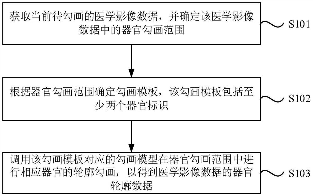
[0021] figure 1 It is a flow chart of the organ contour drawing method provided by Embodiment 1 of the present invention. The technical solution of this embodiment is applicable to the situation of automatically completing the outline drawing of organs of medical image data. The method is preferably executed by a GPU (Graphics Processing Unit, GPU for short) of the medical imaging system provided by the embodiment of the present invention. The medical imaging system includes a CPU (Central Processing Unit, CPU for short) and a GPU, and the two are connected by communication. The method specifically includes the following steps:
[0022] S101. Obtain the current medical image data to be delineated, and determine an organ delineation range in the medical image data.
[0023] Among them, the medical imaging data can be CT (Computed Tomography, referred to as CT, that is, computerized tomography) images, MRI (Magnetic Resonance Imaging, referred to as MRI, nuclear magnetic reso...
Embodiment 2
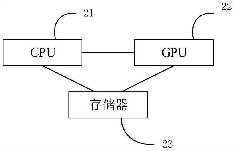
[0050] figure 2 It is a schematic diagram of the medical imaging system provided by Embodiment 2 of the present invention. The system includes a CPU 21, a GPU 22 and a storage device 23; the storage device 23 is used to store one or more first programs and one or more second programs; when the one or more first programs are executed by the CPU 21, so that The CPU 21 acquires medical image data from the corresponding imaging device; when the one or more second programs are executed by the GPU 22, the GPU 22 executes the organ contour delineation method described in the foregoing embodiments.
[0051] Wherein, the imaging equipment may be medical imaging equipment such as CT equipment, MRI equipment, ultrasound equipment, and X equipment.
[0052] In this embodiment, when the GPU detects that there is currently an idle thread, it sends an image acquisition request to the CPU, and when the CPU detects the image acquisition request, it sends the medical image data currently in t...
Embodiment 3
[0077] An embodiment of the present invention also provides a storage medium containing computer-executable instructions, the computer-executable instructions are used to execute a method for contouring an organ when executed by a computer processor, the method comprising:
[0078] Obtain the medical image data currently to be delineated, and determine the delineation range of organs in the medical image data;
[0079] Determining a delineation template according to the organ delineation range, where the delineation template includes at least two organ identifiers;
[0080] Invoking the delineation model corresponding to the delineation template to delineate the contour of the corresponding organ in the delineation range of the organ, so as to obtain the organ contour data of the medical image data.
PUM
 Abstract
Description
Claims
Application Information
 - R&D Engineer
- R&D Manager
- IP Professional
- Industry Leading Data Capabilities
- Powerful AI technology
- Patent DNA Extraction
Browse by: Latest US Patents, China's latest patents, Technical Efficacy Thesaurus, Application Domain, Technology Topic, Popular Technical Reports.
© 2024 PatSnap. All rights reserved.Legal|Privacy policy|Modern Slavery Act Transparency Statement|Sitemap|About US| Contact US: help@patsnap.com