A kind of anti-new coronavirus sars-cov-2 neutralizing nanobody and its application

A nanobody and coronavirus technology, applied in the direction of antiviral agent, virus/bacteriophage, antiviral immunoglobulin, etc., can solve the problem that there is no report of neutralizing nanobody against the new coronavirus SARS-CoV-2

Active Publication Date: 2022-05-17
INST OF PATHOGEN BIOLOGY CHINESE ACADEMY OF MEDICAL SCI
View PDF5 Cites 0 Cited by
  • Summary
  • Abstract
  • Description
  • Claims
  • Application Information

AI Technical Summary

Problems solved by technology

[0005] Currently, there are no reports of neutralizing nanobodies against the novel coronavirus SARS-CoV-2

Method used


Image

  • A kind of anti-new coronavirus sars-cov-2 neutralizing nanobody and its application
  • A kind of anti-new coronavirus sars-cov-2 neutralizing nanobody and its application
  • A kind of anti-new coronavirus sars-cov-2 neutralizing nanobody and its application

Examples


Embodiment 1

[0079] Example 1: Screening of nanobodies recognizing the RBD domain of the SARS-CoV-2 spike protein

[0080] Step 1: Rescue the phage surface-displayed nanobody library

[0081] The fully synthetic nanobody library is stored in the host bacteria in the form of phagemids. Before the panning process begins, the library is rescued to make it a phage-displayed antibody library. The specific method is as follows:

[0082] Take 1mL (OD 600 =100) antibody library Glycerol bacteria, inoculated into 5 bottles of 200mL 2TY-CARB medium (OD 600 =0.1), 37°C, 250rpm shake to OD 600 = about 0.5; add 1.6×10 12 PFU M13KO7, let stand at 37°C for 30 minutes, shake the bacteria at 37°C at 200rpm for 30 minutes; transfer the bacterial liquid to a centrifuge bottle, centrifuge at 2200g for 15 minutes, resuspend the bacteria with 400mL 2TY-CARB-KAN, 30°C, 250rpm Shake the bacteria for 14-16 hours; divide the bacteria solution into centrifuge bottles (200mL per bottle), centrifuge at 10000g, 4°...

Embodiment 2

[0091] Example 2: Fusion expression of nanobody and Fc protein

[0092] Step 1: Construction of recombinant expression vector

[0093] The positive cloned gene was amplified using the upstream primer shown in SEQ ID No.8 and the downstream primer shown in SEQ ID No.9.

[0094] SEQ ID No.8: 5'-ggcgctagccaagttcaattggttgaat-3'

[0095] SEQ ID No.9:

[0096] 5'-tgagcctccactgaattcagaagaaacagtaacttgagtacct-3'.

[0097] A nucleic acid molecule encoding an anti-RBD Nanobody is obtained. The antibody gene was digested with NheI and EcoRI, inserted into the antibody expression vector pCDNA4-Fc, and transformed into DH5a Escherichia coli competent cells (purchased from Quanshijin Company) to obtain recombinant bacteria. The recombinant bacteria were inoculated in liquid LB-Amp medium, cultivated overnight at 37°C, and the plasmid was extracted with a plasmid extraction kit. 16 nanobody recombinant plasmids were obtained. pCDNA4-Fc was transformed from pCDNA4 / myc-HisA (purchased fro...

Embodiment 3

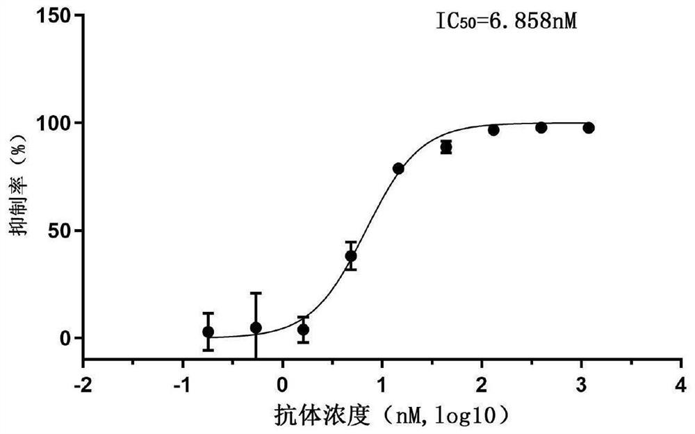
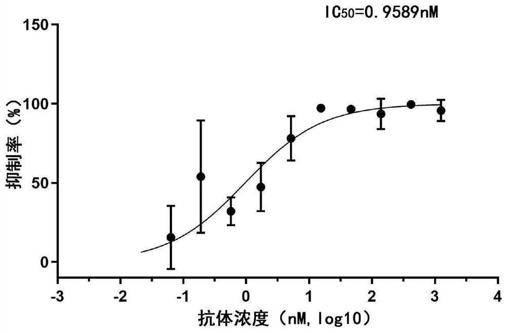
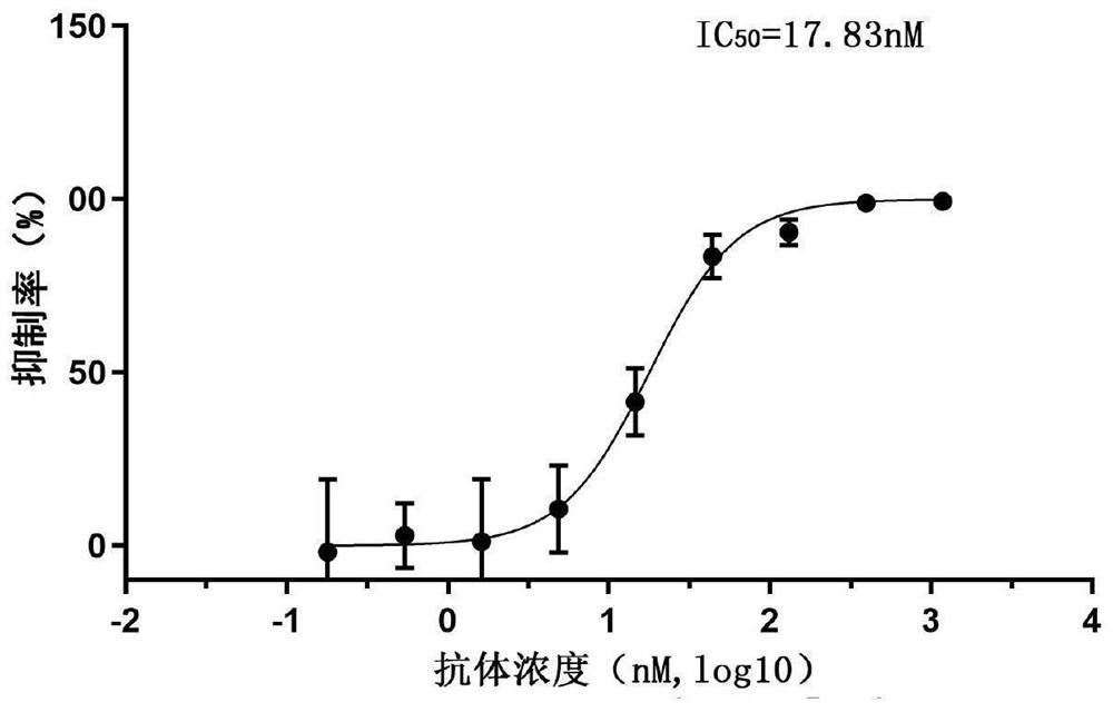
[0100] Example 3: SARS-CoV-2 pseudotyped virus neutralizing activity assay of Nanobodies

[0101] Step 1: SARS-CoV-2 pseudotyped virus packaging

[0102] 6×10 6 HEK293T cells (from the Cell Resource Center, Institute of Basic Medical Sciences, Chinese Academy of Medical Sciences) were inoculated in a 10cm culture dish, and the medium used was DMEM high-glucose medium (purchased from Thermo Fisher) containing 10% fetal bovine serum (purchased from Thermo Fisher). From Thermo Fisher), after 12 hours, transfect 10 μg S gene expression plasmid (Jinweizhi) and 10 μg pNL4.3-Luc-R-E-plasmid (BioVectorNTCC), and replace with 2% fetal bovine serum 12 hours after transfection DMEM high-glucose medium, continue to cultivate for 48 hours and harvest the culture supernatant containing SARS-CoV-2 pseudotyped virus, aliquot and freeze at -80°C.

[0103] Step 2: SARS-CoV-2 pseudotype virus invasion inhibition experiment

[0104] 10 4 Calu-3 cells (from the Cell Resource Center, Institute ...


PUM

PropertyMeasurementUnit
diameteraaaaaaaaaa

Abstract

The invention discloses a neutralizing nanobody against novel coronavirus SARS‑CoV‑2 and an application thereof, belonging to the technical field of biomedicine. The variable region of the neutralizing nanobody has three complementarity determining regions CDR1, CDR2 and CDR3; the neutralizing nanobody is any one of (a1)-(a6). The neutralizing nanobody against novel coronavirus SARS-CoV-2 provided by the present invention can effectively combine with RBD antigen, and can effectively compete with human ACE2 protein for binding to RBD antigen after fusion with human IgG Fc label, and can effectively bind to SARS-CoV-2 pseudotyped virus The high neutralizing activity has important scientific significance and application prospects for the prevention, clinical treatment and development of diagnostic reagents for diseases caused by the new coronavirus SARS‑CoV‑2.

Description

technical field [0001] The invention relates to a neutralizing nanobody against novel coronavirus SARS-CoV-2 and an application thereof, belonging to the technical field of biomedicine. Background technique [0002] The novel coronavirus, known as severe acute respiratory syndrome (SARS)-coronavirus-2 (SARS-CoV-2), is the causative agent of novel coronavirus pneumonia (COVID-19). SARS-CoV-2 uses angiotensin converting enzyme 2 (Angiotensin converting enzyme 2, ACE-2) as a receptor to invade cells and cause lung damage. Respiratory distress syndrome (Acuterespiratory distress syndrome, ARDS) and septic shock, eventually multiple organ failure. Since the outbreak of the epidemic, scientists from all over the world have begun to work on the development of vaccines, antiviral drugs, and antibody drugs. [0003] Antibodies are glycoproteins produced by B cells that proliferate and differentiate into plasma cells after being stimulated by antigens. They can specifically bind to ...

Claims


Application Information

Patent Timeline
Patent Type & Authority Patents(China)
IPC IPC(8): C07K16/10C07K19/00C12N15/85C12N5/10A61K39/42A61P31/14G01N33/569
CPCC07K16/10C12N15/85C12N5/0686A61P31/14G01N33/56983C07K2317/569C07K2317/76C07K2317/565C07K2317/567C07K2319/30C12N2800/107C12N2510/02G01N2333/165G01N2469/10
Inventor 赵振东黄鹤朱悦王蓓王健伟
Owner INST OF PATHOGEN BIOLOGY CHINESE ACADEMY OF MEDICAL SCI
Why Eureka
Try Eureka