A kind of anti-Nipah virus envelope glycoprotein monoclonal antibody and its application
A monoclonal antibody, envelope glycoprotein technology, applied in the field of peptides, to achieve the effect of excellent antigen binding activity
- Summary
- Abstract
- Description
- Claims
- Application Information
AI Technical Summary
Problems solved by technology
Method used
Image
Examples
Embodiment 1
[0024]Example 1. Antibody preparation
[0025]1. Immunize animals
[0026]1) The purified NIV-GP recombinant protein was mixed with the same amount of Freund's complete adjuvant, and injected into the groin of each mouse at multiple points. The injected protein was measured at 20 μg per mouse, and 3 mice were immunized.
[0027]2) After 2 weeks and 4 weeks, the PSCA recombinant protein and Freund's incomplete adjuvant mixture were injected, and the method and dosage were the same as the first time.
[0028]3) Two weeks after the third immunization, the tail vein blood of the mice was taken to test the immunization titer, which reached more than 1:1600 to prepare for fusion, and boosted immunization (20 μg / mouse) 3 days before fusion.
[0029]2. Cell fusion and cloning
[0030]1) Aseptically harvest the mouse spleen, prepare a spleen cell suspension, fuse with Sp2 / 0 myeloma cells according to a conventional method, and culture with HAT selective medium.
[0031]2) On the 4th day after fusion, replace the...
Embodiment 2
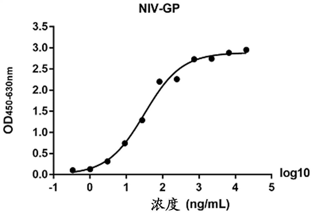
[0050]Example 2. ELISA experiment to detect the binding activity of 14F8 monoclonal antibody and Nipah virus G protein
[0051]1) One day before the experiment, a 96-well ELISA plate was coated with 1μg / mL NIV-GP, and each well was coated with 200μL. Put the coated ELISA plate into a humid box at 4°C overnight.
[0052]2) Wash 5 times with a plate washer on the day of the experiment.
[0053]3) Add 100 μL of blocking solution to each well, and place it at room temperature for 1 hour.
[0054]4) Machine wash the plate 3 times.
[0055]5) Add 150 μL of 14F8 monoclonal antibody at a concentration of 20 μg / mL to the first well, and add 100 μL of diluent to the remaining wells. Aspirate 50μL from the first well and add it to the second well, and so on, with a 1:3 gradient, and the final volume of each well is 100μL. Let stand at room temperature for 1 hour.
[0056]6) Machine wash the plate 5 times.
[0057]7) The HPR-labeled goat anti-mouse IgG secondary antibody was diluted 1:10000 with the diluent, 100 μL...
Embodiment 3
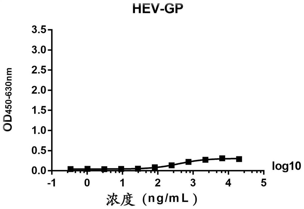
[0062]Example 3. ELISA experiment to detect the binding activity of 14F8 monoclonal antibody to Hendra virus envelope glycoprotein
[0063]1) One day before the experiment, a 96-well ELISA plate was coated with 1μg / mL Hendra virus G protein HEV-GP, and each well was coated with 200μL. Put the coated ELISA plate into a humid box at 4°C overnight.
[0064]2) Wash 5 times with a plate washer on the day of the experiment.
[0065]3) Add 100 μL of blocking solution to each well, and place it at room temperature for 1 hour.
[0066]4) Machine wash the plate 3 times.
[0067]5) Add 150 μL of 14F8 monoclonal antibody at a concentration of 20 μg / mL to the first well, and add 100 μL of diluent to the remaining wells. Aspirate 50μL from the first well and add it to the second well, and so on, with a 1:3 gradient, and the final volume of each well is 100μL. Let stand at room temperature for 1 hour.
[0068]6) Machine wash the plate 5 times.
[0069]7) The HPR-labeled goat anti-mouse IgG secondary antibody was dilut...
PUM
 Abstract
Description
Claims
Application Information
 - R&D
- Intellectual Property
- Life Sciences
- Materials
- Tech Scout
- Unparalleled Data Quality
- Higher Quality Content
- 60% Fewer Hallucinations
Browse by: Latest US Patents, China's latest patents, Technical Efficacy Thesaurus, Application Domain, Technology Topic, Popular Technical Reports.
© 2025 PatSnap. All rights reserved.Legal|Privacy policy|Modern Slavery Act Transparency Statement|Sitemap|About US| Contact US: help@patsnap.com


