Head hierarchical dissection three-dimensional scanning specimen manufacturing method

A technology of three-dimensional scanning and specimen making

Pending Publication Date: 2021-11-09
河南中博科技有限公司
View PDF6 Cites 1 Cited by
  • Summary
  • Abstract
  • Description
  • Claims
  • Application Information

AI Technical Summary

Problems solved by technology

However, the digital 3D specimen of head anatomy has simple structure and complicated process, and there is a certain deviation between the produced specimen and the real specimen, which cannot realize the display of the name of the real structure of the specimen; a single head anatomy 3D scanning specimen can only Create a hierarchical structure, only a single layer of structure can be learned, and it cannot be learned coherently

Method used


Image

  • Head hierarchical dissection three-dimensional scanning specimen manufacturing method
  • Head hierarchical dissection three-dimensional scanning specimen manufacturing method

Examples


Embodiment

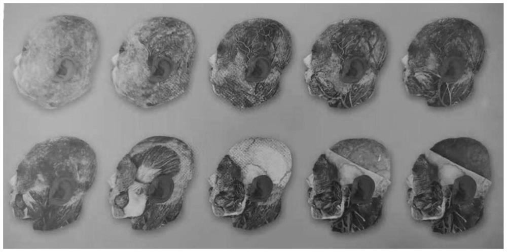
[0048] Example: such as Figure 1-Figure 2 As shown, a method for preparing a head-level anatomical three-dimensional scanning specimen comprises the following steps:

[0049] S1: Select human specimens that are complete, without trauma, without fractures, and without lesions; and perfuse the blood vessels of the specimens, in which the veins are perfused with a mixed solution of blue pigment and latex; after that, 15% formalin is passed through the large blood vessels Infiltrated into various parts of the specimen; after 7 days of parking, arterial perfusion was performed;

[0050] S2: Specimen dissection: including the following steps for hierarchical dissection, and a 3D scan for each dissected layer:

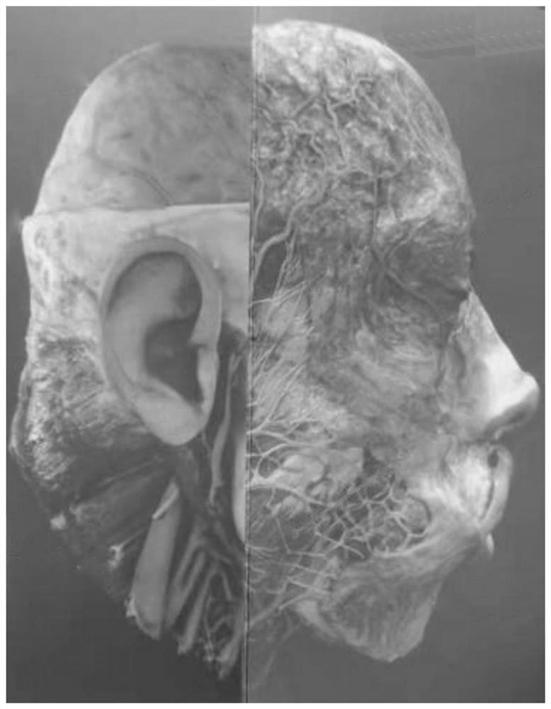
[0051] 1) Remove the skin and show the superficial structures in the superficial fascia, where the superficial structures include the superficial temporal vein, superficial temporal artery and retroauricular vein;

[0052] 2) Remove superficial fascia, reveal galeal aponeu...


PUM


Abstract

The invention relates to a method for manufacturing a head hierarchical dissection three-dimensional scanning specimen. The method comprises the following steps: selecting materials; sequentially removing skin, superficial fascia, latissimus jugular muscle, parotid gland, superficial vascular nerve, cap aponeurosis, masseter, periosteum and temporal muscle, opening zygomatic arch and mandible, removing sternoclavicular mastoid muscle, parietal bone of cranial top, frontal bone, temporal bone and occipital bone, cutting to open superior sagittal sinus, removing dura mater, brain, mandible, zygomatic major muscle, zygomatic minor muscle, orbiculus oculi muscle, trapezius muscle, capsid muscle, diabdominal muscle, deorbital horn muscle cheekbone, endocranium, and veins, removing styloid process tongue bone muscles, external rectus, arteries, styloid process pharynx muscles and styloid process tongue muscles, removing auricles, opening temporal bones, and performing median sagittal incision; then, trimming and cleaning the dissected specimen, and pasting specimen muscles, blood vessels and nerves to the original corresponding positions; and finally, combining and processing the 3D scanned images into a complete digital 3D model, so that the scanned specimen is complete in shape and structure, free switching can be realized, and observation and learning are facilitated.

Description

technical field [0001] The invention relates to the field of preparation of head scanning specimens, in particular to a preparation method of head level anatomical three-dimensional scanning specimens. Background technique [0002] Specimens are animals, plants, minerals, etc., which have been treated in various ways so that they can be preserved for a long time and keep their original appearance as much as possible, so as to provide them for exhibition, demonstration, education, identification, research and other research purposes. Human body specimens are specimens made from real human bodies after a series of chemical treatments. Each human body specimen is a real carrier, containing a large amount of information about individuals and groups. Human anatomy is the most intuitive in anatomy teaching. Teaching aids are the most direct basis for anatomy research. [0003] Regional anatomy is based on systematic anatomy, artificially dividing the human body into eight major p...

Claims


Application Information

Patent Timeline
Patent Type & Authority Applications(China)
IPC IPC(8): G09B23/28
CPCG09B23/28
Inventor 程明亮赵顺贠跃进马万青邢现锋
Owner 河南中博科技有限公司
Why Eureka
Try Eureka