Method, device and system for obtaining brain CT perfusion imaging parameter graph and computer storage medium

A technology of perfusion imaging and parameter map, which is applied in the cross-field of image processing and medical engineering, can solve the problems of low accuracy of perfusion parameter map, high difficulty of perfusion parameter map, and low time resolution of residual function, so as to reduce the difficulty of solving and improve Usability, effect of improving accuracy

Pending Publication Date: 2021-12-28
HANGZHOU ARTERYFLOW TECH CO LTD
View PDF0 Cites 4 Cited by
  • Summary
  • Abstract
  • Description
  • Claims
  • Application Information

AI Technical Summary

Problems solved by technology

[0006] In order to solve the problems of low accuracy of the perfusion parameter map in the prior art, great difficulty in solving the perfusion paramete

Method used


Image

  • Method, device and system for obtaining brain CT perfusion imaging parameter graph and computer storage medium
  • Method, device and system for obtaining brain CT perfusion imaging parameter graph and computer storage medium
  • Method, device and system for obtaining brain CT perfusion imaging parameter graph and computer storage medium

Examples


Embodiment Construction

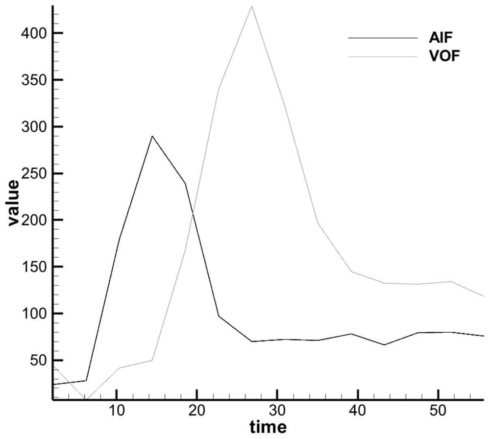
[0057] In the prior art, the deconvolution method is currently the most commonly used CT perfusion image processing method. Refer to the formula (1): c(t)=AIF(t)*k(t); wherein, C(t) is the tissue time-density curve; AIF(t) is the arterial input function; k(t) is the residual function, The specific descriptions of the three belong to common parameters in the prior art, and will not be repeated here. The deconvolution method assumes that the arterial input function AIF(t), the tissue time-density curve C(t) and the residual function k(t) are related to each other through the convolution model, so through the deconvolution operation, the residual function k(t) can be estimated and associated perfusion parameters.

[0058] Since the acquisition time of each frame of CT perfusion image is discrete, it is necessary to discretize the above formula to obtain formula (2):

[0059] in,

[0060] t j is the jth acquisition moment of CT perfusion image;

[0061] c(t j ) for t j Mo...


PUM


Abstract

The invention discloses a method, a device and a system for obtaining a brain CT perfusion imaging parameter graph and a computer storage medium. The method comprises the following steps: obtaining a CT perfusion image, carrying out the pretreatment of the CT perfusion image, and obtaining a discrete tissue density change curve C (n); reading collection time information of the CT perfusion image to obtain an collection time array T (n); intercepting an collection time array T (n) to obtain a relative collection time array t (n); combining the tissue density change curve C (n) with the corresponding relative collection time array t (n) to obtain a tissue density time curve C (tn) of each pixel point; and after fitting or interpolating the discrete tissue density time curve C (tn), re-dispersing according to the same time interval to obtain a discrete tissue density time curve C (n)'; and performing the same processing on the artery input change curve AIF (n) and the vein output change curve VOF (n). According to the method, the availability of the tissue density time curve is improved, the solving difficulty is reduced, and the solving accuracy is improved.

Description

technical field [0001] The present application relates to the interdisciplinary technical field of image processing and medical engineering, in particular to a method, device, system and computer storage medium for obtaining brain CT perfusion imaging parameter maps. Background technique [0002] Stroke, namely stroke, is an acute cerebrovascular disease. It is a group of diseases that cause brain tissue damage due to sudden rupture of blood vessels in the brain or blockage of blood vessels that prevent blood from flowing into the brain, including ischemic and hemorrhagic strokes. High morbidity and high mortality. Changes in the structure of human life have led to an increasing incidence of acute ischemic stroke. Acute ischemic stroke (cerebral infarction) is the most common type of stroke, accounting for 60% to 80% of all strokes. [0003] Perfusion imaging is most widely used in acute stroke and oncology. In stroke diagnosis, the goal of perfusion imaging is to define ...

Claims


Application Information

Patent Timeline
IPC IPC(8): G06T7/00G06T3/40A61B6/03A61B6/00
CPCG06T7/0012G06T3/4007A61B6/032A61B6/481A61B6/501A61B6/507A61B6/5211G06T2207/10081G06T2207/30016G06T2207/10076G06T7/0016G06T2207/30104
Inventor 刘欣何京松史文飞冷晓畅向建平
Owner HANGZHOU ARTERYFLOW TECH CO LTD
Why Eureka
Try Eureka