
Human tissue cavity modeling device and method

A technology of human tissue and modeling method, applied in the field of medical devices, can solve the problems of not considering the elasticity and volume of the inner wall of vaginal tissue, increasing the risk of inserting a needle into the bladder and rectum, increasing the pain of patients and the risk of bleeding infection, etc. The effect of reducing bleeding and infection risk, facilitating image segmentation, and reducing the chance of occurrence

Active Publication Date: 2021-12-31
ZHONGNAN HOSPITAL OF WUHAN UNIV
View PDF19 Cites 0 Cited by
  • Summary
  • Abstract
  • Description
  • Claims
  • Application Information

AI Technical Summary

Problems solved by technology

[0003] Studies have shown that at this stage, based on fillings such as gauze and cylinders, the elasticity and volume of the inner wall of the vaginal tissue are not considered. The volume and position of the filling are manually judged based on human experience, and five problems are prone to occur: 1) Insufficient vaginal filling (gauze ), there is a lack of filling (cylindrical cylinder) in the fornix, resulting in tissue gaps between the model and the vagina and the inner wall of the fornix, which will lead to underdosage of the tumor and affect the accuracy of dose calculation; One side and farther away, insufficient filling of the cervix and vagina will lead to more normal tissues (such as the vaginal wall) that the needle passes through, increasing the pain of the patient and the risk of bleeding infection; 3) Since the vagina is adjacent to the bladder and rectum, insufficient filling will also lead to inability to Keep the bladder and rectum away from the needle channel, increasing the risk of the needle breaking the bladder and rectum; 4) Too much filling, resulting in a model that is large inside and small outside, and because of the limited elasticity of the inner wall of the vaginal entrance, it will cause discomfort to the patient during treatment ;5) Since the fillers such as gauze have the same density as soft tissue, it is difficult to distinguish from the inner wall of the vagina on CT images, resulting in inaccurate modeling and affecting the accuracy of the model

Method used


Image

  • Human tissue cavity modeling device and method
  • Human tissue cavity modeling device and method
  • Human tissue cavity modeling device and method

Examples


Embodiment Construction

[0048] The following will clearly and completely describe the technical solutions and specific implementation methods of the present invention with reference to the accompanying drawings in the present invention.

[0049] In order to better understand the present invention, the following will further illustrate the present invention in conjunction with specific examples. It should be understood that these examples are only for illustrating the present invention and do not limit the scope of the present invention. In order to make the object, technical solution and advantages of the present invention clearer, the present invention will be further described in detail below in conjunction with the accompanying drawings and embodiments. It should be understood that the specific embodiments described here are only used to explain the present invention, not to limit the present invention.

[0050] In order to achieve the above purpose, as Figure 1-5As shown, the present invention ...


PUM


Abstract

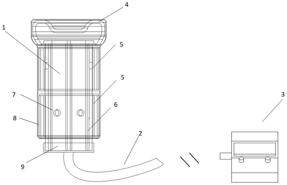
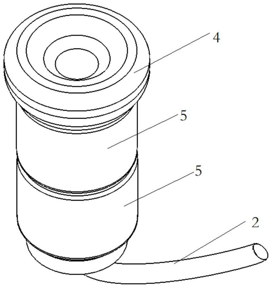
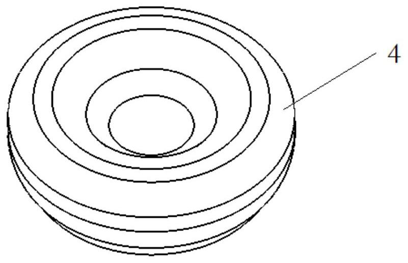
The invention discloses a human tissue cavity modeling device and method. The human tissue cavity modeling device comprises a filling rod, a connecting air hose, a control host, an air pump and an electromagnetic valve; the filling rod comprises a top concave air bag, an annular air bag and a multi-branch-passage air pipe; the multi-branch-passage air pipe is sequentially communicated with the connecting air hose and the air pump; and the electromagnetic valve is controlled by the control host to be opened and closed to independently inflate each air bag of the inflation rod. An inflation volume and air bag diameter function is constructed through an in-vitro experiment, the widths of different positions of the vagina of a patient are measured, modeling is carried out based on a direct insertion needle model, the optimal inflation volume of each air bag is determined, after inflation is completed in vivo, CT scanning is carried out on the patient with the filling rod, and the filling rod is automatically segmented for modeling according to different CT values of air and tissues. According to the invention, the vagina and the fornix can be perfectly filled according to the individualized vagina width, the defect of traditional fornix filling is overcome, the normal tissue needle inserting distance is reduced, the safety, comfort and dosage precision of needle insertion are improved, and pain and bleeding infection risks are reduced.

Description

technical field [0001] The invention relates to the technical field of medical devices, in particular to a human tissue cavity modeling device and method. Background technique [0002] There are many complex tissue cavities in the human anatomy, such as the external auditory canal, nasopharyngeal cavity, oral cavity, female reproductive system, etc. High-precision modeling of tissue cavities has important clinical application value, such as surgical path planning, surgical plan simulation, brachytherapy pin phantom design, etc. Taking brachytherapy as an example, due to the specific location and shape of tumor stumps in some patients with cervical cancer, the traditional dosimetry system cannot meet the requirements of dose distribution. In order to achieve personalized optimal dosimetry distribution, some researchers are currently using 3D printing technology to make implant phantoms, which greatly improves the dose distribution. The most important step in the production ...

Claims


Application Information

Patent Timeline
Patent Type & Authority Applications(China)
IPC IPC(8): A61B34/10
CPCA61B34/10A61B2034/105
Inventor 沈九零陈敏邱惠王骁踊刘晖谢丛华张俊陈纪
Owner ZHONGNAN HOSPITAL OF WUHAN UNIV
Learn More