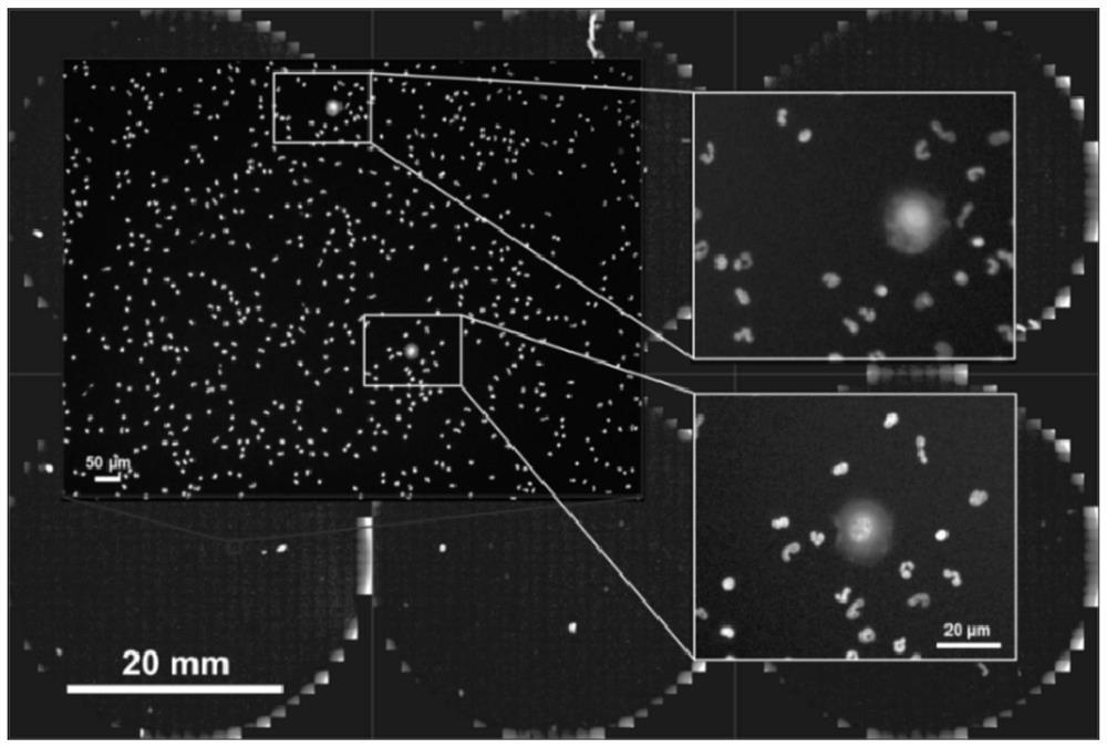
Specific method for detecting breast cancer circulating tumor cells by adopting HER2 antibody immunofluorescence method
A tumor cell, immunofluorescence technology, applied in the field of molecular biology testing, to avoid false negative results, accurate results, and increase the difficulty of work
- Summary
- Abstract
- Description
- Claims
- Application Information
AI Technical Summary
Problems solved by technology
Method used
Image
Examples
Embodiment 1
[0027] A specific method for detecting circulating tumor cells in breast cancer by using HER2 antibody immunofluorescence method, comprising the following steps: S1: collecting 8ml of peripheral blood from patients with breast cancer, mixing the collected blood sample with lysate at a ratio of 1:8, and mixing at room temperature 15min. Centrifuge at 200RCF for 5 minutes, aspirate the supernatant and keep the cells. Add 2mL of 1% FPBS to the centrifuge tube, mix well and centrifuge at 200RCF for 5 minutes, remove the supernatant; add 1ml of FPBS and mix well, add the mixed cells into the treated culture dish, and incubate at 37°C for 45min After culturing, put the petri dish in a 4°C refrigerator and let it stand for 10 minutes; suck off the FPBS, add 4% formaldehyde to the petri dish and put it at 4°C for 10 minutes. Remove formaldehyde by suction, add 1mL of methanol, and place at -20°C for 10min. Aspirate off the methanol and wash three times with 2 mL of PBS each time.
...
Embodiment 2
[0045] A specific method for detecting circulating tumor cells in breast cancer by using HER2 antibody immunofluorescence method, comprising the following steps: S1: collecting 9ml of peripheral blood from patients with breast cancer, mixing the collected blood sample with lysate at a ratio of 1:8, and mixing at room temperature 15min. Centrifuge at 200RCF for 5 minutes, aspirate the supernatant and keep the cells. Add 2mL of 1% FPBS to the centrifuge tube, mix well and centrifuge at 200RCF for 5 minutes, remove the supernatant; add 1ml of FPBS and mix well, add the mixed cells into the treated culture dish, and incubate at 37°C for 45min After culturing, put the petri dish in a 4°C refrigerator and let it stand for 10 minutes; suck off the FPBS, add 4% formaldehyde to the petri dish and put it at 4°C for 10 minutes. Remove formaldehyde by suction, add 1mL of methanol, and place at -20°C for 10min. Aspirate off the methanol and wash three times with 2 mL of PBS each time.
...
Embodiment 3
[0063] A specific method for detecting circulating tumor cells in breast cancer by using HER2 antibody immunofluorescence method, comprising the following steps: S1: collecting 10 ml of peripheral blood from patients with breast cancer, mixing the collected blood sample with lysate at a ratio of 1:9, and mixing at room temperature 15min. Centrifuge at 200RCF for 5 minutes, aspirate the supernatant and keep the cells. Add 2mL of 1% FPBS to the centrifuge tube, mix well and centrifuge at 200RCF for 5 minutes, remove the supernatant; add 1ml of FPBS and mix well, add the mixed cells into the treated culture dish, and incubate at 37°C for 45min After culturing, put the petri dish in a 4°C refrigerator and let it stand for 10 minutes; suck off the FPBS, add 4% formaldehyde to the petri dish and put it at 4°C for 10 minutes. Remove formaldehyde by suction, add 1mL of methanol, and place at -20°C for 10min. Aspirate off the methanol and wash three times with 2 mL of PBS each time. ...
PUM
 Abstract
Description
Claims
Application Information
 - R&D
- Intellectual Property
- Life Sciences
- Materials
- Tech Scout
- Unparalleled Data Quality
- Higher Quality Content
- 60% Fewer Hallucinations
Browse by: Latest US Patents, China's latest patents, Technical Efficacy Thesaurus, Application Domain, Technology Topic, Popular Technical Reports.
© 2025 PatSnap. All rights reserved.Legal|Privacy policy|Modern Slavery Act Transparency Statement|Sitemap|About US| Contact US: help@patsnap.com

