Intravascular ultrasound image processing method based on deep learning

A deep learning, ultrasound image technology, applied in the field of image processing, can solve the problems of large impact, low contrast and low resolution of intravascular ultrasound images, achieve accurate automatic identification and evaluation analysis, improve display clarity, reduce redundant parts Effect

Pending Publication Date: 2022-02-08
孟令波
View PDF0 Cites 2 Cited by
  • Summary
  • Abstract
  • Description
  • Claims
  • Application Information

AI Technical Summary

Problems solved by technology

In the medical field, especially in pathological and radiological film reading, surgical robots, etc., intelligent diagnosis functions have begun to take shape, but there are many limiting factors in the field of ultrasound: due to the lack of uniform standards for the collection of ultrasound images, image quality Acquisition is restricted by the doctor's operation, and different angles and different acquisition positions have a great influence on the results. At the same time, different tissue structures in ultrasound images vary greatly, and the transmission of dynamic images during acquisition has higher requirements for the recognition and diagnosis of artificial intelligence images. AI development for ultrasound work is still in its infancy
[0003] Through the above analysis, the problems and defects of the existing technology are: the contrast of the existing intravascular ultrasound image is low, it is greatly affected by noise and various artifacts, it is difficult to distinguish the tissue characteristics of the plaque; the resolution is low, Cause different readers to draw different conclusions

Method used


Image

  • Intravascular ultrasound image processing method based on deep learning
  • Intravascular ultrasound image processing method based on deep learning
  • Intravascular ultrasound image processing method based on deep learning

Examples


Embodiment Construction

[0047]In order to make the object, technical solution and advantages of the present invention more clear, the present invention will be further described in detail below in conjunction with the examples. It should be understood that the specific embodiments described here are only used to explain the present invention, not to limit the present invention.

[0048] Aiming at the problems existing in the prior art, the present invention provides a method for processing intravascular ultrasound images based on deep learning. The present invention will be described in detail below with reference to the accompanying drawings.

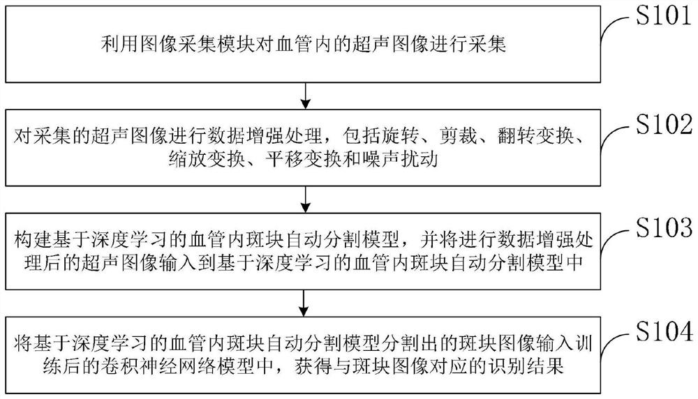
[0049] Such as figure 1 As shown, the deep learning-based intravascular ultrasound image processing method provided by the embodiment of the present invention includes:

[0050] S101, using the image acquisition module to acquire ultrasound images in blood vessels;

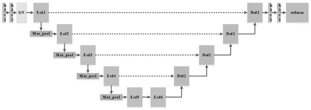
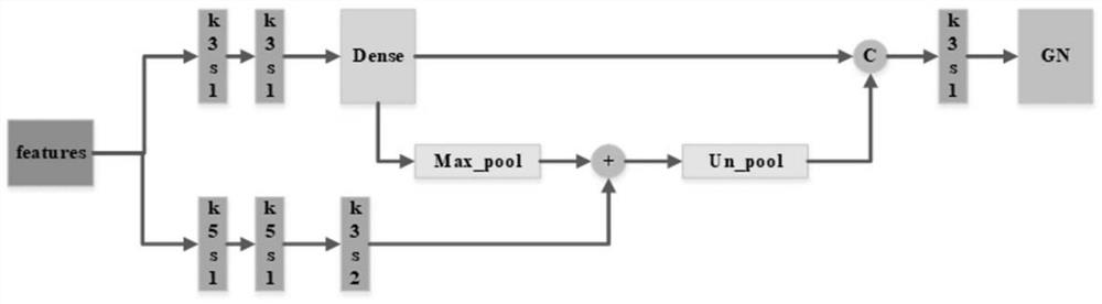
[0051] S102, performing data enhancement processing on the collected ultrasound image, incl...


PUM


Abstract

The invention belongs to the technical field of image processing, and discloses an intravascular ultrasonic image processing method based on deep learning, which comprises the following steps of: performing data enhancement processing on an collected ultrasonic image, and inputting the ultrasonic image subjected to the data enhancement processing into an intravascular plaque automatic segmentation model based on deep learning, and inputting the plaque image segmented by the automatic segmentation model into the trained convolutional neural network model to obtain an recognition result corresponding to the plaque image. According to the method, the display definition of the ultrasonic image can be effectively improved by performing data enhancement processing on the collected ultrasonic image, the redundant part of the image is reduced, the size of the image is simplified, the intravascular plaque image can be automatically segmented accurately and quickly through the intravascular plaque automatic segmentation model based on deep learning, and the accuracy of the intravascular plaque segmentation is improved. The method facilitates the subsequent automatic recognition through the convolutional neural network model, and achieves the quick, objective and accurate automatic recognition and evaluation analysis of the vascular lesion.

Description

technical field [0001] The invention belongs to the technical field of image processing, and in particular relates to an intravascular ultrasound image processing method based on deep learning. Background technique [0002] At present: intravascular ultrasound (IVUS) is the most direct and accurate method for diagnosing coronary heart disease at present. According to the differences in the acoustic characteristics of different vascular tissue components, different ultrasonic echo signals are obtained, so as to obtain a real-time tomographic image of the two-dimensional cross-section of the blood vessel including the blood vessel wall structure and plaque tissue, and realize the detection of blood vessels. Quantitative assessment of mural and intimal atherosclerotic lesions. However, due to the low contrast of ultrasound images, which are greatly affected by noise and various artifacts, it is still difficult to distinguish the tissue characteristics of plaques. In addition,...

Claims


Application Information

Patent Timeline
Patent Type & Authority Applications(China)
IPC IPC(8): G06T7/00G06T7/11G06T5/00G06N3/04G06N3/08
CPCG06T7/0012G06T7/11G06T5/002G06N3/08G06T2207/10132G06T2207/20021G06T2207/20081G06T2207/20084G06T2207/30101G06N3/045
Inventor 孟令波张超张健
Owner 孟令波
Why Eureka
Try Eureka