Collection device for resected specimen under digestive endoscopy

A collection device and digestive endoscope technology, which is applied in the field of medical devices, can solve problems such as damage, inability to preserve specimen tissue, and affect the storage time and quality of specimen tissue, and achieve the effect of preventing impact

Inactive Publication Date: 2022-02-11
MUDANJIANG MEDICAL UNIV
View PDF1 Cites 0 Cited by
  • Summary
  • Abstract
  • Description
  • Claims
  • Application Information

AI Technical Summary

Problems solved by technology

[0005] When the above-mentioned sample collection device collects the sample tissue by using negative pressure, the sample tissue is easily damaged when it moves under the action of negative pressure, thereby affecting the later pathological analysis. At the same time, the above-mentioned sample collection device cannot effectively collect the sample tissue. Preservation cannot guarantee the activity of the specimen tissue, which affects the storage time and quality of the specimen tissue;

Method used


Image

  • Collection device for resected specimen under digestive endoscopy
  • Collection device for resected specimen under digestive endoscopy
  • Collection device for resected specimen under digestive endoscopy

Examples


Embodiment 1

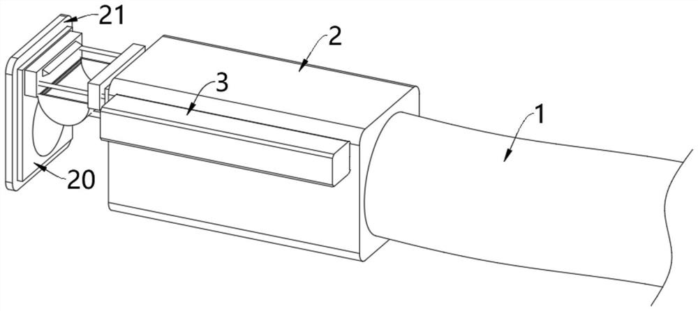
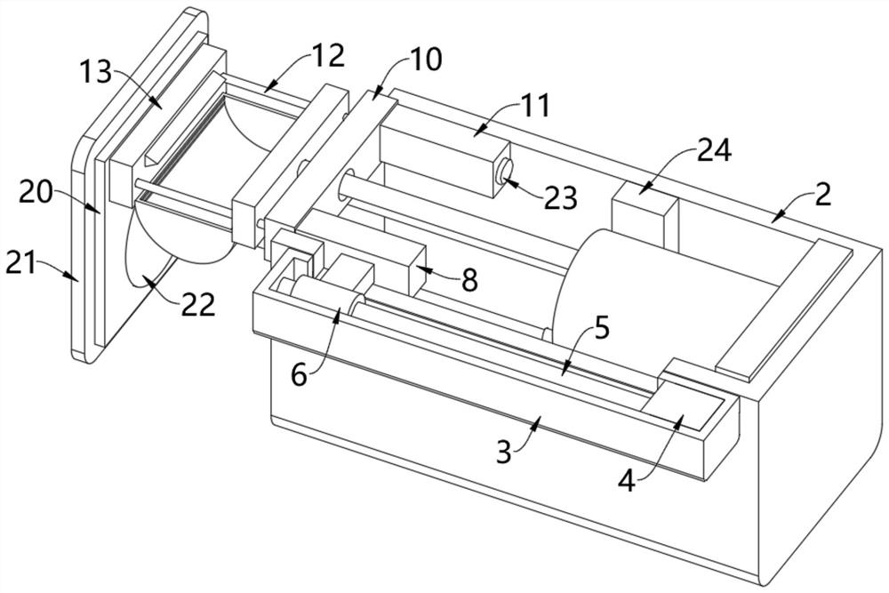
[0043] see Figure 1-Figure 6 , a device for collecting resection specimens under digestive endoscopy, comprising an insertion tube 1, an outer cylinder 2 is fixedly installed at the end of the insertion tube 1, one end of the outer cylinder 2 is open, and a connecting plate 10 is slidably arranged on the inner wall of the opening end of the outer cylinder 2, The left and right ends of the right side of the connecting plate 10 are respectively fixed with a first slide plate 8 and a second slide plate 11, and both the first slide plate 8 and the second slide plate 11 are slidably arranged on the inner wall of the outer cylinder 2;

[0044] The outer wall of the outer cylinder 2 is fixed with a side cylinder 3, the inner wall of the side cylinder 3 is fixed with a motor 4, the top of the output shaft of the motor 4 is fixed with a screw 5, and the screw 5 is rotated inside the side cylinder 3. The outer wall of 5 is threadedly connected with a screw nut 6, and the outer wall of ...

Embodiment 2

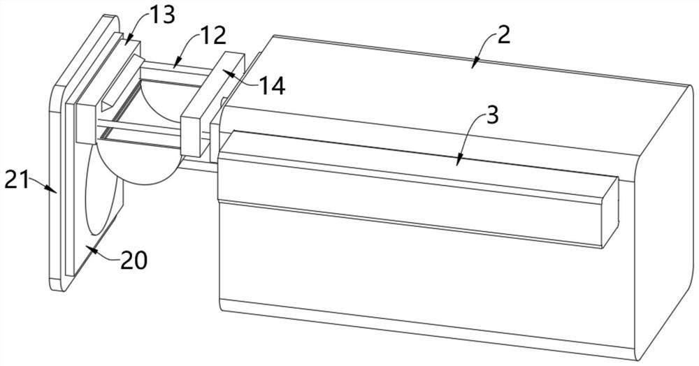
[0051] see Figure 7 , on the basis of Embodiment 1, a sealing plate 20 matching the opening end of the outer cylinder 2 is fixedly installed on the outer wall of the first clamping block 13 facing away from the second clamping block 14, and the left side of the sealing plate 20 is fixed The cover plate 21 is installed, and after the first clamping block 13 gradually approaches the second clamping block 14 and cuts off the specimen tissue, continue to control the first clamping block 13 to move to the right, and the first clamping block 13 continues to move to the right to make its left side The sealing plate 20 and the cover plate 21 cover the opening end of the outer cylinder 2 to seal the outer cylinder 2, thereby storing and storing the whole mechanism and preventing the external environment from affecting the specimen tissue;

[0052] The inside of the cover plate 21 is provided with a semiconductor refrigeration chip 22, and the end of the second slide plate 11 is provid...

Embodiment 3

[0055] see Figure 8 , on the basis of Embodiment 1 or Embodiment 2, a push rod 25 is fixedly installed in the middle of the outer wall on the right side of the second clamping block 14, and the end of the push rod 25 passes through the connecting plate 10 and is fixedly connected with a piston 28, and the piston 28 moves Set on the inner wall of the storage cavity 29, the storage cavity 29 is opened inside the inner cylinder 26 and the tail plate 27, and the tail plate 27 is fixedly installed on the right inner wall of the outer cylinder 2;

[0056] The inside of the storage cavity 29 is stored with RNAlater solution, which can protect the RNA in the sample tissue from being degraded. In the RNAlater solution, the RNA of the sample tissue can be stored stably for one day at 37 degrees Celsius. It can be stored for seven days at 25 degrees Celsius, four weeks at two to eight degrees Celsius, and permanent at minus 20 degrees Celsius. The usage of RNAlater solution is also very...


PUM


Abstract

The invention relates to the technical field of medical instruments, and discloses a digestive endoscopy resection specimen collecting device. The device comprises an insertion tube, an outer cylinder is fixedly mounted at the tail end of the insertion tube, one end of the outer cylinder is open, and a connecting plate is slidably arranged on the inner wall of the open end of the outer cylinder; a first sliding plate and a second sliding plate are fixedly mounted at the left and right ends of the right side of the connecting plate; after the first clamping block gradually gets close to the second clamping block and cuts off the specimen tissue, the first clamping block is continuously controlled to move rightwards, at the moment, the first clamping block also pushes the second clamping block to move rightwards, the second clamping block moves rightwards and can push the piston to move rightwards through the push rod, the piston moves rightwards to push and extrude the RNAlater solution in the storage cavity, the RNAlater solution is extruded and can be conveyed into the collecting bag through the conveying pipe, the first one-way valve and the connecting hose, so that the specimen tissue is soaked, the activity of the specimen tissue is guaranteed, the storage time of the specimen tissue is prolonged, and the storage quality of the specimen tissue is improved.

Description

technical field [0001] The invention relates to the technical field of medical devices, in particular to a device for collecting resection specimens under digestive endoscopy. Background technique [0002] Endoscopic treatment techniques involve early resection of superficial digestive tract malignant tumors under endoscopy, debulking treatment of some advanced malignant tumors, and gastrointestinal lesions that cannot be clearly diagnosed by endoscopic biopsy. With the development of science and technology, digestive endoscopy technology has made great progress, and the design of the specimen recovery device under digestive endoscopy is becoming more and more ingenious, and its functions are becoming more and more abundant; [0003] For example, the Chinese patent announcement number is: CN108403155A invention patent, which just provides a kind of resection specimen collection device under digestive endoscopy, including a base, the left side of the base is rotatably connect...

Claims


Application Information

Patent Timeline
Patent Type & Authority Applications(China)
IPC IPC(8): A61B17/00A61B17/3209A61B10/04A61B10/00
CPCA61B17/00234A61B17/3209A61B17/320016A61B10/04A61B10/0096A61B2017/00287
Inventor 刘秀玉张红梅马占荣柳凤芝都伟王景波佟玥玥陶英歌陈燕陈兵陈翠戴毓
Owner MUDANJIANG MEDICAL UNIV
Why Eureka
Try Eureka