
Renal fibrosis evaluation method based on magnetic resonance elastography and serological examination

A technique for renal fibrosis and fibrinogen, which is applied in the field of renal fibrosis assessment based on magnetic resonance elastography and serological examination, can solve problems such as a stable renal fibrosis assessment method, and achieves simple operation and good stability. , the effect of high sensitivity

Pending Publication Date: 2022-02-22
SHUGUANG HOSPITAL AFFILIATED WITH SHANGHAI UNIV OF T C M
View PDF2 Cites 0 Cited by
  • Summary
  • Abstract
  • Description
  • Claims
  • Application Information

AI Technical Summary

Problems solved by technology

[0004] However, there is no method for assessing renal fibrosis based on magnetic resonance elastography and serological examination with high sensitivity and good stability as described in this application

Method used


Image

  • Renal fibrosis evaluation method based on magnetic resonance elastography and serological examination
  • Renal fibrosis evaluation method based on magnetic resonance elastography and serological examination
  • Renal fibrosis evaluation method based on magnetic resonance elastography and serological examination

Examples


Embodiment 1

[0019] 1. MRE inspection

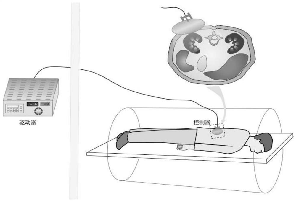
[0020] MRI system ( figure 1 ): Shanghai United Imaging Medical Technology Co., Ltd. uMR 780; the multifunctional portable magnetic resonance elastography excitation device is self-made, in which the excitation device generates shear waves by electromagnetic drive, including two parts, the controller and the driver. The controller is placed in the scanning The outside of the room is used to generate low-frequency shear waves. The driver passes through the wall through the plastic connecting tube and enters the scanning room. The front end is made of custom-made circular silicone material that can fully fit the chest and abdomen wall of the human body, and will generate uniform and soft sound while ensuring comfort. shear wave.

[0021] Empty urine before the examination, the patient prone position. The coil selection is a 32-channel body phased array surface coil, and the body surface positioning is marked as the spinocostal angle. The scanning ran...

Embodiment 2

[0030] This embodiment provides a kit for evaluating renal fibrosis, consisting of:

[0031] Three biomarkers (fibrinogen, fibrinogen degradation products, and D-dimer) and their enzyme-labeled antibodies, carbonate buffer at pH 9.6, phosphate buffer at pH 7.4, serum protein Diluent, stop solution, tetramethylbenzidine substrate solution.

[0032] It further includes an instruction manual, which describes the following:

[0033] 1. Perform MRE to detect the kidney elasticity value of the individual;

[0034] 2. Detection of individual serum fibrinogen, fibrinogen degradation products and D-dimer levels;

[0035] 3. Substitute the above test results into the equation: n=0.285MRE+0.835 fibrinogen+1.05 fibrinogen degradation product+0.41D dimer+3.413, and calculate the value of n which represents the degree of renal fibrosis.


PUM


Abstract

The invention relates to a renal fibrosis evaluation method based on magnetic resonance elastography and serological examination, which specifically comprises the following steps: performing magnetic resonance elastography examination and serum biomarker fibrinogen, fibrinogen degradation product and D dimer examination on an individual, and substituting into an evaluation model: n = 0.285MRE + 0.835 fibrinogen + 1.05 fibrinogen degradation product + 0.41 D dimer + 3.413, and calculating to obtain a renal fibrosis degree evaluation result. The method disclosed by the invention is novel, high in sensitivity, good in stability, simple and convenient to operate and noninvasive.

Description

technical field [0001] The invention relates to the technical field of pathological diagnosis, in particular to a method for evaluating renal fibrosis based on magnetic resonance elastography and serological examination. Background technique [0002] Renal fibrosis is a common pathway of chronic kidney disease leading to end-stage renal failure. Early detection of renal fibrosis and accurate assessment of the degree of renal fibrosis play an important role in early clinical intervention and selection of appropriate treatment methods. However, there is currently a lack of reliable, noninvasive, and quantitative methods for evaluating renal fibrosis. Needle biopsy is still the gold standard for the diagnosis of renal fibrosis. However, needle biopsy has few materials, large errors, and is an invasive operation, which is not well accepted by patients. Therefore, exploring new non-invasive methods for assessing renal fibrosis has important clinical value. Ultrasound elastogra...

Claims


Application Information

Patent Timeline
IPC IPC(8): A61B5/20G01N33/68A61B5/055
CPCA61B5/055A61B5/201G01N33/6893G01N2800/347
Inventor 杨雪军袁杰冯原戴勇鸣詹松华
Owner SHUGUANG HOSPITAL AFFILIATED WITH SHANGHAI UNIV OF T C M
Learn More