
Patsnap Eureka
For R&D, Patsnap Eureka makes reading and utilizing patents & technical documents easy.
Patsnap Eureka AIR
Designed for self-driven R&D workflows. Generate viable solutions, solve complex R&D challenges, empower your innovation with AI.

Patsnap Eureka Materials
Designed for material experts only. Revolutionize your material R&D, from search, analyze, to developing new materials.

TechResearch
Generate reliable direction feasibility study reports for your R&D in just a few steps.

TechSeek
Discover and master advanced knowledge NOW. Basics, ideas, possibilities, all at once.

TechMind
As an expert in R&D Theories, TechMind can generates customized viable solutions instantly.

TechRisk
Analyze your overall solution with one click, know your potential R&D risks in advance.

TechMonitor
Get weekly tech updates, stay abreast of the latest tech innovations and key insights.
Temporomandibular joint positioning device for laboratory mouse
A temporomandibular joint and positioning device technology, which is applied in the fields of animal restraint equipment, medical science, veterinary equipment, etc., can solve the problems of unreliable experimental results, injury of experimental mice, and low experimental efficiency, so as to improve experimental efficiency and The effect of relieving pain and reducing the difficulty of operation
- Summary
- Abstract
- Description
- Claims
- Application Information
AI Technical Summary
Problems solved by technology
Method used
Image

Examples
Embodiment 1
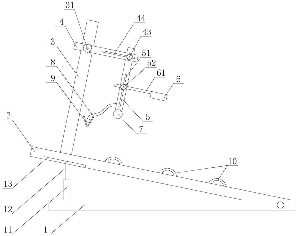
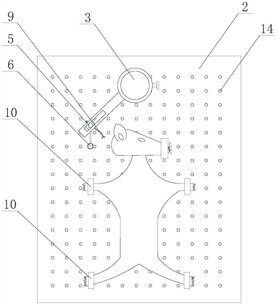
[0048] like Figure 2 to Figure 5 The shown temporomandibular joint positioning device for experimental mice includes a working board 2, on which a fixture 10 for fixing an experimental mouse is arranged, and on said working board 2, a supporting column 3 is provided, and said supporting column 3 Connect the ear hole positioning part 5, the ear hole positioning part 5 is used to insert the external auditory canal of the experimental mouse and snap into the ear hole 21 of the experimental mouse, the ear hole positioning part 5 is provided with a connecting bar 8, and the connecting bar 8 is connected There is a buckle 9 for snapping to the root of the zygomatic arch 23 of the experimental rat.
[0049] In this embodiment, the operation difficulty can be reduced by combining the work board, which is suitable for novices. When the workload is heavy or skilled hands are required to complete the operation quickly, the ear hole positioning member 5, connecting strip 8 and buckle 9 ...
Embodiment 2
[0058] On the basis of Example 1, such as Figure 2 to Figure 5 As shown, the ear hole positioning member 5 is provided with a third sliding groove 53, the guide ring handle 61 can move in the third sliding groove 53, and the third sliding groove 53 is connected with a third locking groove 51 , the third locking groove 51 is provided with a third locking member 52, and the third locking member 52 is used to fix the position of the guide ring handle 61 in the third slide groove 53; The support column 3 is provided with a cantilever 4, and the cantilever 4 is provided with a collar 41 for being sleeved on the support column 3. The collar 41 is threaded with a first locking member 31. A locking member (31) is used to fix the position of the collar 41 on the support column 3; the cantilever 4 is provided with a second chute 42, and the ear hole positioning member 5 can be positioned in the second chute 42, the second chute 42 is communicated with a second locking groove 44, and a...
Embodiment 3
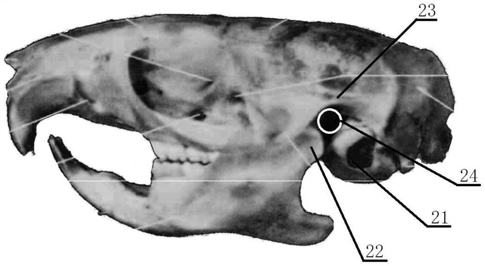
[0063] On the basis of the above-mentioned embodiments, a guide ring handle 61 is provided on the ear hole positioning member 5, and an injection guide ring 6 is connected to the guide ring handle 61, and the bottom of the injection guide ring 6 is used for embedding the experimental 24 in the joint cavity of the rat.
[0064] In one or more embodiments, the bottom end of the injection guide extends down and beyond the guide handle to form a raised portion 69 .
[0065] In this example, if figure 1 with Figure 9 As shown, after the positions of the ear hole positioning member 5 and the buckle 9 are fixed, the position of the depressed joint cavity can be quickly and conveniently determined by using the bottom of the injection guide ring 6, that is, the position between the condyle 22, the root of the zygomatic arch 23, and the ear hole 21. area, and can directly embed the bottom of the injection guide ring 6 into the joint cavity 24 to form a fixed point, thereby forming a ...
PUM
 Abstract
Description
Claims
Application Information
 - R&D Engineer
- R&D Manager
- IP Professional
- Industry Leading Data Capabilities
- Powerful AI technology
- Patent DNA Extraction
Browse by: Latest US Patents, China's latest patents, Technical Efficacy Thesaurus, Application Domain, Technology Topic, Popular Technical Reports.
© 2024 PatSnap. All rights reserved.Legal|Privacy policy|Modern Slavery Act Transparency Statement|Sitemap|About US| Contact US: help@patsnap.com