Ophthalmology simulation teaching equipment and method

An ophthalmology and equipment technology, applied in the field of binocular indirect ophthalmoscopy, can solve problems such as the inability to see the fundus, and achieve the effects of facilitating the simulation of scleral top pressure, improving the adaptability of use, and improving the practicality of use.

Active Publication Date: 2022-05-24
SHENZHEN MATERNITY & CHILD HEALTHCARE HOSPITAL +1
View PDF5 Cites 1 Cited by
  • Summary
  • Abstract
  • Description
  • Claims
  • Application Information

AI Technical Summary

Problems solved by technology

The invention can solve the problem that the examiner needs to coordinate hand-eye-head position. During the examination, the examiner's pupil-eyepiece-objective lens-patient's pupil must be on the same straight line, otherwise the fundus cannot be seen, and the inspection of the peripheral retina needs to cooperate with the top of the sclera. In order to see the situation of the peripheral retina clearly, it is necessary to know the exact pressing position, pressing direction and pressing force.

Method used


Image

  • Ophthalmology simulation teaching equipment and method
  • Ophthalmology simulation teaching equipment and method
  • Ophthalmology simulation teaching equipment and method

Examples


Embodiment 1

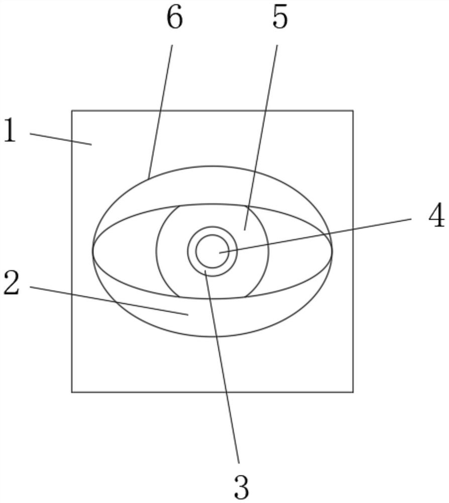
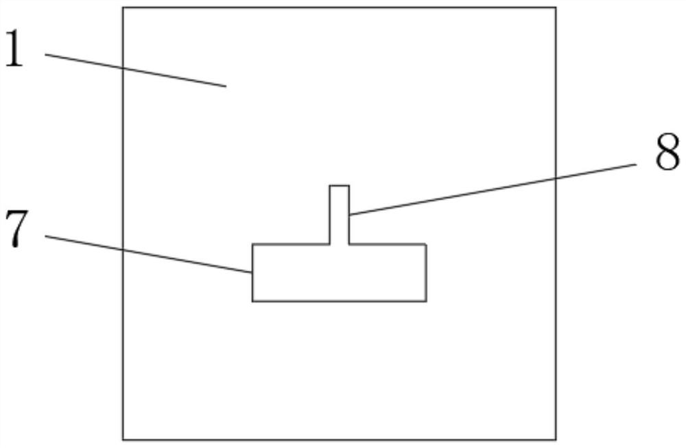
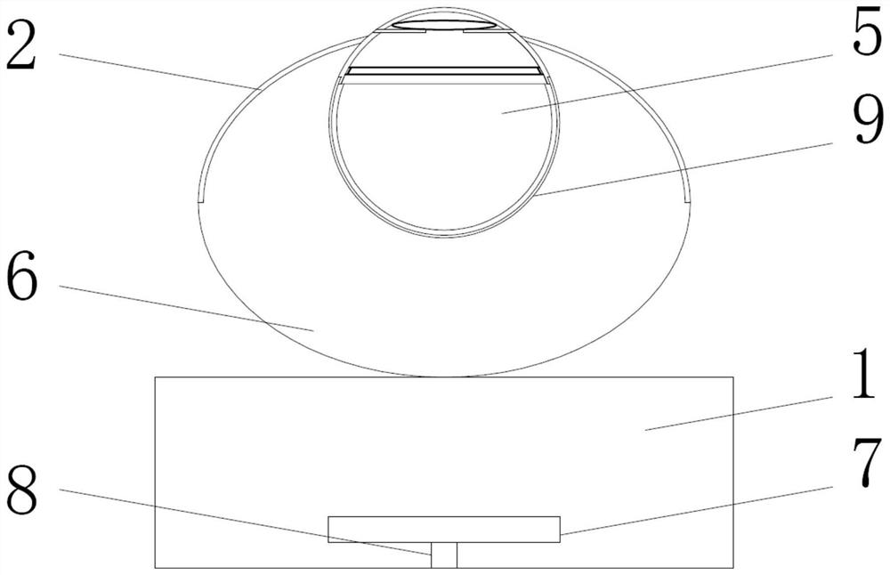
[0044] like Figure 1 to Figure 5 As shown, in order to achieve the above purpose, the present invention provides the following technical solutions: a kind of ophthalmic simulation teaching equipment, comprising a base 1 and a main body 6, a main body 6 is installed in the middle of the front surface of the base 1, and a rotation groove 9 is arranged in the middle of the front surface of the main body 6, The eyeball model 5 is installed on the inner surface of the rotation groove 9, the eyelid model 2 is installed on the front surface of the main body 6 at the top and the bottom of the eyeball model 5, the lower hemisphere 12 is installed inside the eyeball model 5 near the rear surface, and the inner surface of the lower hemisphere 12 is installed with the fundus. Picture 11, the inside of the eyeball model 5 is installed with the upper hemisphere 10 near the front surface through the connecting platform 13, the upper hemisphere 10 and the lower hemisphere 12 are elastic rubbe...

Embodiment 2

[0055] like Figure 1 to Figure 5 As shown in the figure, an ophthalmology simulation teaching equipment proposed by the present invention, compared with the first embodiment, the present embodiment further includes: the light shielding sheet 3 is provided with a circular middle hole, and the light shielding sheet 3 is provided with a total of five , the diameters of the middle holes of the five shading sheets 3 are respectively 4cm, 5cm, 6cm, 7cm and 8cm, which is convenient for simulating the pupil size after mydriasis of common infants and adults in clinical practice.

Embodiment 3

[0057] In the ophthalmology simulation teaching equipment proposed by the present invention, compared with the first embodiment, the rotating groove 9 and the eyeball model 5 in this embodiment are installed with interference fit, so as to prevent the eyeball model 5 from Rotation without external force affects the simulation teaching. In this embodiment, during use, the 360-degree rotation of the eyeball is achieved through the interference fit of the rotating groove 9 and the eyeball model 5, such as Figure 4 or Figure 7 As shown, the user can select different lenses 4 to simulate the refraction state of the eyes of infants and adults, which is common in clinical practice, and teach students. Students wear binocular indirect ophthalmoscope on their head. On this simulated teaching equipment, learn and repeat the practice of adjusting the relative distance and angle of hand-eye-head, and adjusting the position and strength of scleral pressure until they can check and recor...


PUM


Abstract

The invention provides an ophthalmology simulation teaching device and method, the ophthalmology simulation teaching device comprises a base and a main body, the main body is installed in the middle of the front surface of the base, a rotating groove is formed in the middle of the front surface of the main body, and an eyeball model is installed on the inner surface of the rotating groove; eyelid models are mounted on the front surface of the main body and at the top and the bottom of the eyeball model, a lower hemisphere is mounted in the eyeball model close to the rear surface, an upper hemisphere is mounted in the eyeball model close to the front surface through a connecting table, and a lens is mounted in the middle of the front surface of the upper hemisphere; an anti-dazzling screen is installed on the rear surface of the lens in the upper hemisphere, a sawtooth edge picture is installed on the inner surface of the upper hemisphere and behind the lens, and a fundus picture is installed on the inner surface of the lower hemisphere. The invention provides the ophthalmology simulation teaching equipment which has the characteristics of simulating human eyelid tissues, eyeball structures, diopter ranges, different pupil sizes and the like, and a more vivid simulation teaching basis can be provided for ophthalmologists and medical students.

Description

technical field [0001] The invention relates to the technical field of binocular indirect ophthalmoscope inspection, in particular to an ophthalmology simulation teaching device; and further provides an ophthalmology simulation teaching method using the ophthalmology simulation teaching device. Background technique [0002] Binocular indirect ophthalmoscopy is an important means of clinical fundus disease screening, diagnosis and treatment, and it is a skill that ophthalmologists must master. Compared with direct ophthalmoscopy, binocular indirect ophthalmoscopy has the advantages of three-dimensional sense, large visible range, clear imaging, and less influence by refractive media. If used with a scleral depressor, the periphery of the retina can be inspected. Binocular indirect ophthalmoscopy is currently the gold standard for retinopathy of prematurity screening. The main treatment method for severe ROP, retinal laser photocoagulation under direct vision with binocular ...

Claims


Application Information

Patent Timeline
Patent Type & Authority Applications(China)
IPC IPC(8): G09B23/28
CPCG09B23/28
Inventor 杨冰芝张国明杨传忠陈懿连朝辉郑棉瑩
Owner SHENZHEN MATERNITY & CHILD HEALTHCARE HOSPITAL
Why Eureka
Try Eureka