
Three-dimensional culture method for in-vitro endometrial gland organs

A technology of endometrium and three-dimensional culture, applied in the field of biomedicine

Pending Publication Date: 2022-05-27
LIAONING GREATEST BIO-PHARM CO LTD
View PDF2 Cites 1 Cited by
  • Summary
  • Abstract
  • Description
  • Claims
  • Application Information

AI Technical Summary

Problems solved by technology

[0007] The purpose of the present invention is to provide a three-dimensional culture method of human endometrial gland organoids, aiming at solving the problem of three-dimensional culture of human endometrial gland organoids in vitro using human endometrial curettage tissue. Questions about culture, subculture, cryopreservation, and recovery methods;

Method used


Image

  • Three-dimensional culture method for in-vitro endometrial gland organs
  • Three-dimensional culture method for in-vitro endometrial gland organs
  • Three-dimensional culture method for in-vitro endometrial gland organs

Examples


Embodiment 1

[0070] Example 1. In vitro three-dimensional culture of endometrial glandular organoids, their cryopreservation and thawing

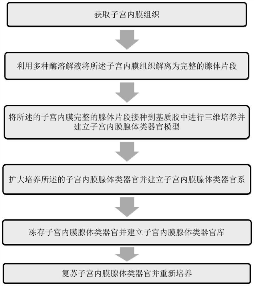
[0071] Fresh endometrial tissue after hysterectomy was obtained from 10 subjects aged 18-38 years old who signed the informed consent form and without other diseases, and the decidua part of the endometrium was taken and placed in phosphate buffer and quickly transferred to sterile laboratory, such as figure 1 The method steps shown are to create a three-dimensional endometrial gland organoid model. Some samples were cryopreserved to establish endometrial gland organoid bank. The specific steps are as follows:

[0072] Sample collection:

[0073] Endometrial tissue sample collection: Fresh endometrial tissue was obtained by hysterectomy.

[0074] Transport of endometrial tissue samples: Place endometrial tissue in phosphate buffered saline, record patient information, seal the tube cap tightly, place the transport container on ice, and quickly trans...

Embodiment 2

[0084] Example 2. Light microscopy, electron microscopy and immunofluorescence detection of endometrial gland organoids

[0085] The present invention is further verified by the following experimental examples for the genetic consistency of the human endometrial gland organoids obtained by the above in vitro method of the present invention and the organs from which they are derived.

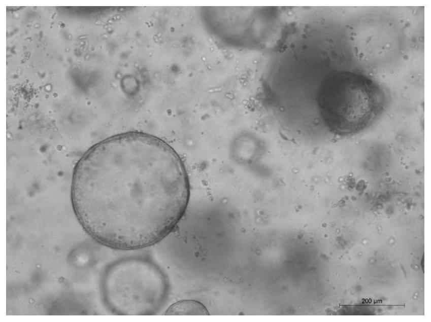
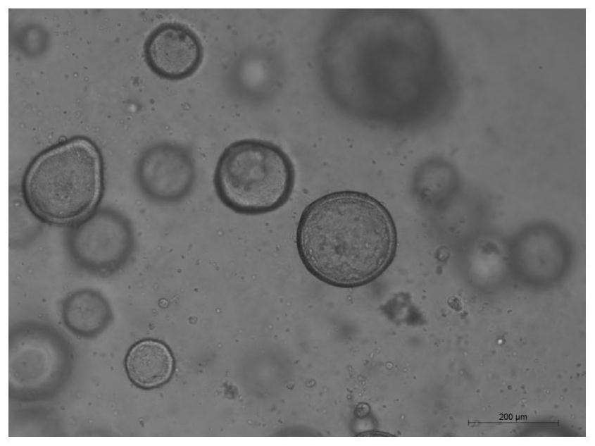
[0086] First, we observed the human endometrial glandular organoids obtained after steps 3), 4) and 6) of Example 1, and after repeating steps 1) to 4) of Example 1 22 times under an optical microscope. Morphology, which was found to be similar to the derived human endometrium (Exemplary light-microscopic morphology of human endometrial glandular organoids obtained after the above steps, respectively Figures 2A-2D middle).

[0087] Under the light microscope, single-layer columnar cells can be seen closely arranged into a glandular lumen-like structure, and secretions and a small amount of exfo...


PUM


Abstract

The invention relates to a three-dimensional culture method for in-vitro endometrial gland organs. Specifically, the invention relates to a three-dimensional culture, passage, freezing and unfreezing method of the in-vitro endometrial gland type organ, the in-vitro endometrial gland type organ obtained by the method and application of the in-vitro endometrial gland type organ.

Description

technical field [0001] The invention belongs to the technical field of biomedicine, and mainly relates to a three-dimensional culture method of human endometrial gland organoids. Background technique [0002] The uterus is a complex organ with an inner endometrium composed of a single layer of pseudostratified luminal epithelium (LE) and branched columnar glandular epithelium (GE), supported by mesenchymal fibroblasts as well as immune cells, blood vessels, and the lymphatic system , these glands are thought to be important for establishing pregnancy through interactions with other endometrial cells and the embryo. Meanwhile, the endometrium is one of the most dynamic tissues in the human body, undergoing a hormone-regulated growth cycle and shedding each month. Evidence suggests that endometrial stem / progenitor cells are responsible for the regenerative properties of the endometrium. [0003] The process of human embryo implantation is a complex physiological process. At ...

Claims


Application Information

Patent Timeline
IPC IPC(8): C12N5/071C12Q1/02A01N1/02
CPCC12N5/0682G01N33/5014A01N1/0221C12N2533/90C12N2509/00C12N2509/10C12N2500/30C12N2500/38C12N2501/119C12N2501/415G01N2500/10
Inventor 张雷张亚楠盖波张轶商微
Owner LIAONING GREATEST BIO-PHARM CO LTD
Learn More