Application of METs as molecular marker in evaluation of clinical prognosis of glioma patient
A molecular marker and glioblastoma technology, applied in the field of medicine, can solve the problems of less than 10% 5-year survival rate of glioma patients, short survival period of patients, glioma invasion and other problems, and improve the overall Survival period, the effect of improving clinical treatment effect
- Summary
- Abstract
- Description
- Claims
- Application Information
AI Technical Summary
Problems solved by technology
Method used
Image
Examples
Embodiment 1
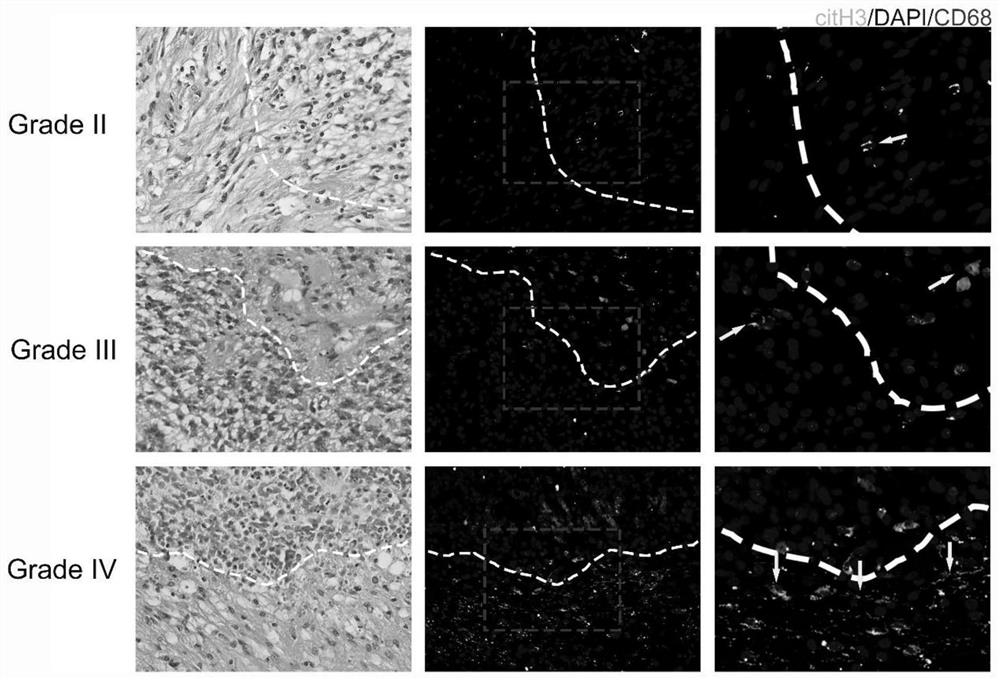
[0029] Example 1 The level of METs around the tumor of brain glioblastoma increased
[0030] 1. Preparation of detection reagents
[0031] (1) Preparation of fluorescent antibody;
[0032] Add 10 mg bovine serum albumin dry powder to a 1.5 mL EP tube, dissolve in 1 mL phosphate buffered saline (PBS, pH=7.4), and add 1 μg CD68 mouse monoclonal antibody and 2 μg citH3 rabbit monoclonal antibody to the EP tube , mixed thoroughly with the pipette, and temporarily stored in a -20°C refrigerator;
[0033] (2) Preparation of fluorescent secondary antibodies;
[0034] Add 10 mg bovine serum albumin dry powder to a 1.5 mL light-proof EP tube, dissolve it in 1 mL PBS, and take 2 μg goat anti-rabbit IgG (H+L) Alexa Fluor 488 and 2 μg goat anti-mouse IgG (H +L) Add Alexa Fluor 594 into a light-proof EP tube, mix thoroughly with a pipette, and temporarily store in a -20°C refrigerator away from light;
[0035] (3) Preparation of DAPI staining agent;
[0036] Add 10mg bovine serum albu...
Embodiment 2
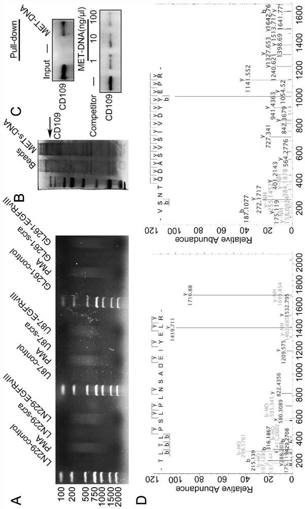
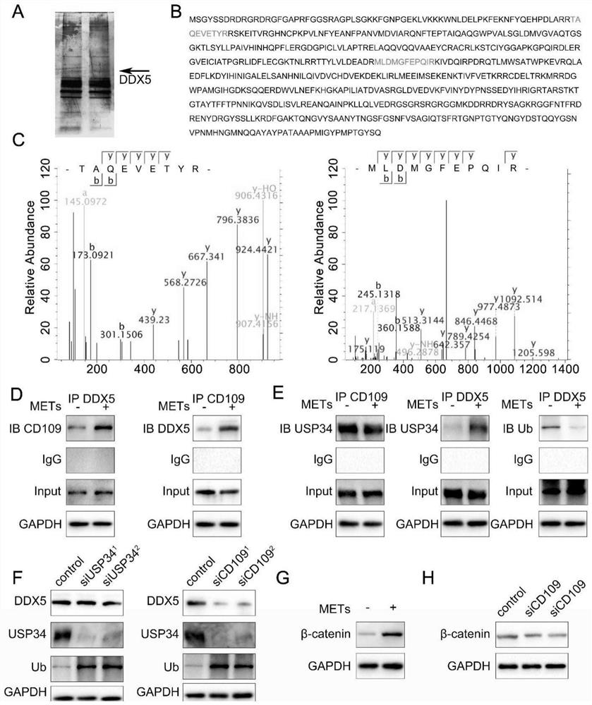
[0060] DNA fragments in Example 2METs bind GBM cell membrane proteins and promote downstream signaling pathways
[0061] 1. Experimental method
[0062] 1.1 DNA extraction and preparation
[0063] (1) Add 500nM phorbol ester (PMA) into the culture medium of LN229 and HG7 cells, put it in the cell incubator and let it stand for 4h, discard the culture medium, add 2mL of pre-cooled PBS solution to wash, and centrifuge at 1000g for 10min to get the supernatant;
[0064] (2) Using a sonicator (power: 50w, sonication: 10s, interval: 30s, a total of 15 cycles) to crush the extracted MET-DNA to obtain DNA fragments with a length of about 200-1000bp.
[0065] 1.2 DNA pull-down experiment
[0066] (1) Mix 5 μg of biotin-labeled DNA and 500 μg of GBM cell membrane protein in advance, and place on ice;
[0067] (2) Take 100 μL of Streptavidin-agarose G beads, wash with 0.5 mL of pre-cooled PBS solution, and centrifuge at 5000 g for 30 seconds;
[0068] (3) Add the mixture of DNA and ...
Embodiment 3
[0109] Example 3 METs can promote the malignant progression of GBM
[0110] 1. Method
[0111] 1.1 By using 5 μg ml of GBM cell line LN229 and primary cell HG7 -1 The cells were treated with METs for 12 hours, and the changes after treatment were observed by Transwell chamber and cell scratch experiment. Stimulate LN229 and HG7 cells with 500nM PMA for 4h, discard the medium, add 2mL cold PBS to wash, centrifuge at 1000g for 10min, and take the supernatant for later use.
[0112] 1.2 Using Transwell technology to detect changes in cell migration and invasiveness in cell lines after adding METs
[0113] (1) Spread Matrigel: Dilute Matrigel gel with serum-free cell culture medium at a ratio of 1:8 at 4°C, take 100 μL and evenly spread it on the surface of the polycarbonate membrane in the upper chamber, and place it at 37°C for 0.5-1h;
[0114] (2) Cell culture: Take the cells to be tested in the logarithmic growth phase, wash with PBS, suspend the cells with serum-free mediu...
PUM
 Abstract
Description
Claims
Application Information
 - R&D
- Intellectual Property
- Life Sciences
- Materials
- Tech Scout
- Unparalleled Data Quality
- Higher Quality Content
- 60% Fewer Hallucinations
Browse by: Latest US Patents, China's latest patents, Technical Efficacy Thesaurus, Application Domain, Technology Topic, Popular Technical Reports.
© 2025 PatSnap. All rights reserved.Legal|Privacy policy|Modern Slavery Act Transparency Statement|Sitemap|About US| Contact US: help@patsnap.com


