Lung CT image segmentation device based on spatial neighborhood analysis

A technology of neighborhood analysis and CT images, applied in the field of image processing, can solve the problems of not fully considering the three-dimensional spatial information of the lesion area, poor interpretability, difficult clinical practice, etc., to achieve low overall calculation, ensure accuracy, and improve segmentation The effect of precision

Pending Publication Date: 2022-05-27
SHANGHAI HANYU BIOLOGICAL SCI & TECH CO LTD
View PDF0 Cites 2 Cited by
  • Summary
  • Abstract
  • Description
  • Claims
  • Application Information

AI Technical Summary

Problems solved by technology

[0004] First, the image segmentation method for a single layer only uses the two-dimensional spatial information of the cross-section of the CT image, which makes it difficult to accurately segment the boundary area of ​​some lesions
The layer thickness of high-resolution CT images is usually about 1 mm, and the actual size of most lesions to be segmented is much larger than the layer thickness. Therefore, it is difficult for a single cross-sectional layer to provide effective information in the other two orthogonal directions in three-dimensional space, resulting in The segmentation results are discontinuous or missing;
[0005] Second, the intra-slice spacing (resolution) of CT images is usually smaller than the inter-slice spacing (slice thickness), that is, there is anisotropy in the orthogonal direction, which leads to the need for image segmentation methods that use three-dimensional space regions as processing units through image interpolation The pixel spacing in all directions is consistent, and different interpolation strategies will directly affect the accuracy of the image segmentation of the lesion area;
[0006] Third, the deep learning method represented by three-dimensional convolution has stronger spatial feature analysis capabilities and is not affected by anisotropy, but its overall parameter scale and calculation volume have increased by an order of magnitude, which has great impact on hardware equipment. High requirements, and the image segmentation method based on 3D supervision information relies on the complete 3D annotation of the lesion area, so it is difficult to apply to clinical practice; Fourth, in the existing medical image processing schemes, there is no specific lung CT lesion The solution to segmenting scenes, and the general deep learning segmentation method uses the image features of the supervisory information (image annotation data) as a guide to achieve result reasoning. Its interpretability for specific lesion areas is poor. How to flexibly apply it to clinical diagnosis Practice and combination with the actual needs of physicians are still issues that need to be considered
[0007] Through literature search, it is found that there is an implicit reverse attention mechanism for segmenting the 2019-nCoV lesion area in chest CT images. This method only targets a single CT layer for lesion area segmentation, and does not fully consider the inter-CT layer The three-dimensional spatial information of the lesion area contained; another method performs two-dimensional convolution on continuous CT layer slices to realize the segmentation of three-dimensional blood vessel structures. This method integrates image features of multiple adjacent two-dimensional CT layer slices in the channel domain , and extract the semantic information of a single layer from the fusion feature through non-local attention, but this scheme only integrates the features of the local neighborhood image in the channel dimension, and fails to truly analyze the single pixel and the three-dimensional spatial neighborhood. Relevant semantic information for other pixels

Method used


Image

  • Lung CT image segmentation device based on spatial neighborhood analysis
  • Lung CT image segmentation device based on spatial neighborhood analysis
  • Lung CT image segmentation device based on spatial neighborhood analysis

Examples


Embodiment Construction

[0055] In order to make the structure and advantages of the present application clearer, the structure of the present application will be further described below with reference to the accompanying drawings.

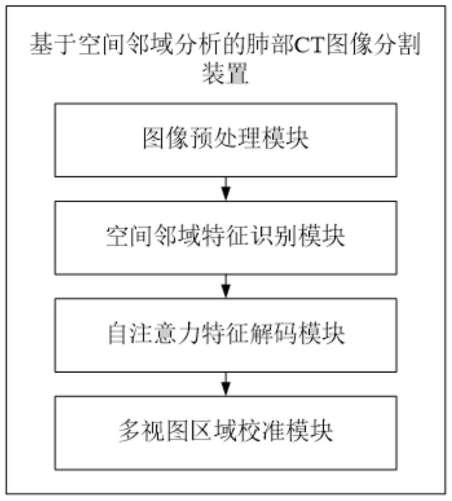
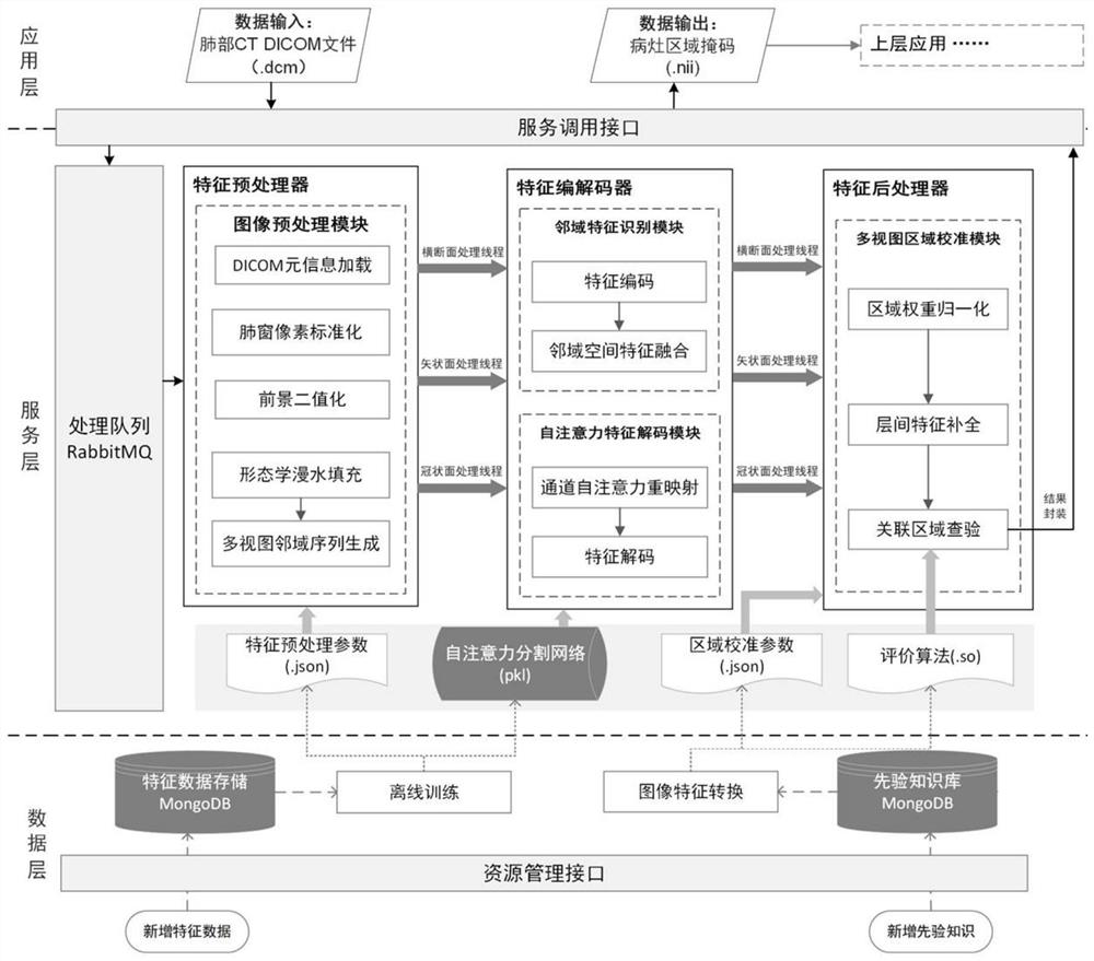
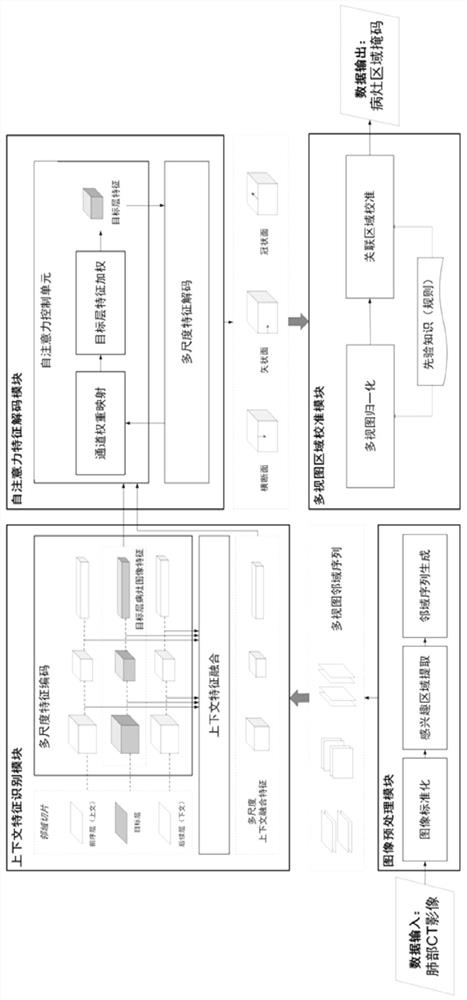
[0056] combined with Figure 1 to Figure 5 , a lung CT image segmentation device based on spatial neighborhood analysis proposed in the embodiment of the present application includes an image preprocessing module, a spatial neighborhood feature identification module, a self-attention feature decoding module, and a multi-view region calibration module, wherein: The image preprocessing module normalizes the CT value under the lung window to the image pixel value according to the original lung CT image file, calculates the lung parenchyma foreground area mask corresponding to each layer, and combines the single foreground area layer and its front and rear neighborhood maps. The layers are combined into a set of neighborhood sequences for the subsequent feature extraction pro...


PUM


Abstract

The invention provides a lung CT image segmentation device based on spatial neighborhood analysis. On the basis of extracting two-dimensional image features of a single CT layer, contextual three-dimensional image features between neighborhood CT sequences are fused in parallel through three-dimensional convolution, and expression of three-dimensional focus area image features is achieved while the full 3D convolution operand and the parameter scale are reduced; meanwhile, channel domain two-dimensional image feature components corresponding to each neighborhood layer slice sequence are remapped from the context fusion feature map by utilizing a self-attention mechanism, so that the feature decoding process of a single CT layer is guided, and the focus image segmentation accuracy is improved; in order to improve the adaptability and interpretability of the algorithm, interpretable priori knowledge is introduced as an additional image segmentation judgment rule, so that the segmentation result is calibrated and checked, and a basis is provided for clinical auxiliary diagnosis.

Description

technical field [0001] The present application relates to the field of image processing, in particular to a lung CT image segmentation device based on spatial neighborhood analysis. Background technique [0002] A lung CT image is a series of cross-sectional sequential image layers obtained by computed tomography. Using image processing technology to locate and segment lung CT lesions, it can provide image visualization and quantitative analysis results for radiologists, and then provide help for clinical diagnosis and disease detection. [0003] Existing lung CT image segmentation devices usually process a single layer, or analyze 3D images composed of all layers, but various processing methods have the following problems: [0004] First, the image segmentation method for a single layer only uses the two-dimensional spatial information of the cross-section of the CT image, which makes it difficult to accurately segment some lesion boundary areas. The layer thickness of hi...

Claims


Application Information

Patent Timeline
Patent Type & Authority Applications(China)
IPC IPC(8): G06T7/11G06K9/62G06N3/04G06V10/80
CPCG06T7/11G06T2207/10081G06T2207/20104G06T2207/30061G06N3/045G06F18/253
Inventor 何玮罗楹王崇宇章曾姜丽红蔡鸿明
Owner SHANGHAI HANYU BIOLOGICAL SCI & TECH CO LTD
Why Eureka
Try Eureka