Fundus image blood vessel segmentation method based on skeleton prior and contrast loss

A fundus image and skeleton technology, applied in the field of medical image processing, can solve problems such as poor performance, achieve the effects of suppressing interference, improving robustness, and promoting learning

Active Publication Date: 2022-05-31
UNIV OF ELECTRONICS SCI & TECH OF CHINA
View PDF17 Cites 3 Cited by
  • Summary
  • Abstract
  • Description
  • Claims
  • Application Information

AI Technical Summary

Problems solved by technology

However, although the existing methods have a good overall effect on blood vessel segme...

Method used


Image

  • Fundus image blood vessel segmentation method based on skeleton prior and contrast loss
  • Fundus image blood vessel segmentation method based on skeleton prior and contrast loss
  • Fundus image blood vessel segmentation method based on skeleton prior and contrast loss

Examples


Embodiment Construction

[0041] In order to facilitate technical personnel to understand the technical content of the present invention, the technical solution of the present invention is further explained below in conjunction with the accompanying drawings.

[0042] Consult the literature in related fields, and download the existing common open source fundus image blood vessel segmentation dataset:

[0043] DRIVE (download address is http: / / www.isi.uu.nl / Research / Databases / DRIVE)

[0044] STARE (download address is http: / / www.ces.clemson.edu / ahoover / stare / probing / index.html)

[0045] CHASE DB1 (download address is http: / / blogs.kingston.ac.uk / retinal / chasedb1 / )

[0046] HRF (download address is http: / / www5.informatik.uni-erlangen.de / research / data / fundus-images)

[0047] Divide each data set into a training set and a test set (according to experience, the division ratio is 1:1), and crop the color fundus images in the training set. The cropping size of each image is 128*128, and the cropping method i...


PUM


Abstract

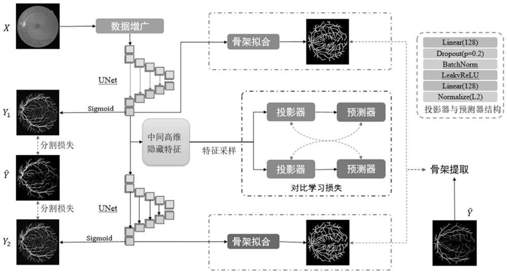
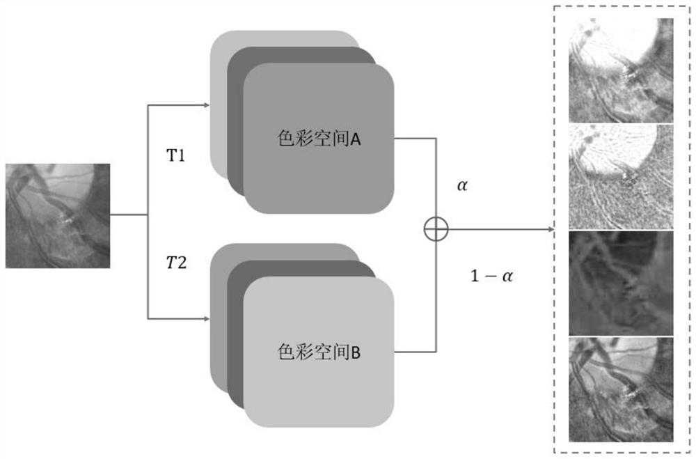
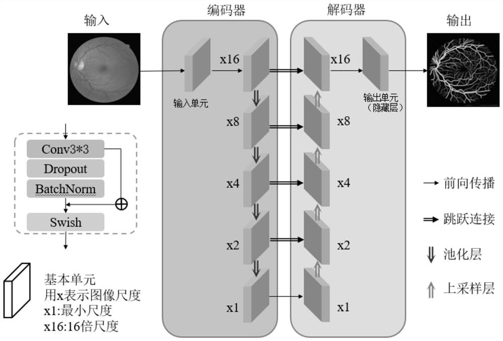
The invention discloses a fundus image blood vessel segmentation method based on skeleton prior and contrast loss. The fundus image blood vessel segmentation method comprises the following steps: S1, performing data augmentation on a color fundus image; s2, performing expert annotation on the eye fundus image to extract a skeleton; s3, inputting the eye fundus image into a segmentation network, and calculating segmentation loss; s4, the foreground and background features of the middle features are compared to learn loss; s5, outputting a skeleton continuity constraint for the segmentation model, and solving a loss function; s6, superposing the three loss functions to obtain total loss, carrying out gradient back propagation, and stopping training when the total loss is not reduced any more for four consecutive rounds; and S7, obtaining a binary vascular tree segmentation result. Compared with the prior art, the contrast loss function adopted in the two types of pixel feature sample sets can further improve the discrimination capability of the model for hidden layer features in a high-dimensional space, can extract small blood vessels and prevent the blood vessels from being broken, can inhibit the interference of biomarkers, and is very suitable for fine retinal vessel tree segmentation.

Description

technical field [0001] The invention belongs to the field of medical image processing, in particular to a blood vessel segmentation method for fundus images based on skeleton prior and contrast loss. Background technique [0002] In the diagnosis of ophthalmic diseases, color fundus images, as a non-invasive means of vascular imaging, have become an important reference material for ophthalmologists due to their convenient acquisition and low cost. The retina contains an extremely rich number of intertwined and scattered blood vessels, and the branches of these retinal blood vessels form a complete vascular tree. The shape of the branches of the vascular tree, including the caliber of the blood vessels near and far from the optic disc, the branch angles of the blood vessel branches, and the degree of curvature of the blood vessels all reflect the health of the human eye system, and can even reflect many other cardiovascular diseases. However, retinal imaging is also affected...

Claims


Application Information

Patent Timeline
IPC IPC(8): G06T7/11G06T7/136G06T7/194G06N3/04G06N3/08G06T5/30G06T5/50
CPCG06T7/11G06T7/136G06T7/194G06T5/30G06T5/50G06N3/084G06T2207/10024G06T2207/20221G06T2207/30101G06T2207/30041G06T2207/20081G06T2207/20084G06N3/048G06N3/045
Inventor 李永杰谭玉博杨开富
Owner UNIV OF ELECTRONICS SCI & TECH OF CHINA
Why Eureka
Try Eureka