
Medical image segmentation method based on highlight spot removal

A medical image and image segmentation technology, applied in the field of image recognition, can solve problems such as incomplete semantic information, blurred images, and inability to meet the requirements of image restoration

Pending Publication Date: 2022-07-05
ANHUI UNIVERSITY
View PDF0 Cites 0 Cited by
  • Summary
  • Abstract
  • Description
  • Claims
  • Application Information

AI Technical Summary

Problems solved by technology

[0008] There are problems such as incomplete semantic information and blurred images in the results of traditional image restoration methods, which cannot meet the current requirements for image restoration

Method used


Image

  • Medical image segmentation method based on highlight spot removal
  • Medical image segmentation method based on highlight spot removal
  • Medical image segmentation method based on highlight spot removal

Examples


Embodiment 1

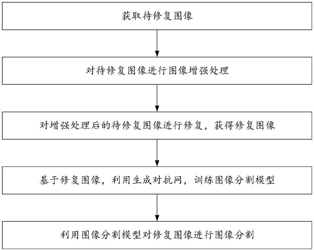
[0053] A medical image segmentation method based on highlight removal, such as figure 1 shown, the specific steps are:

[0054] Step 1. Obtain the image to be repaired;

[0055] Step 2, performing image enhancement processing on the image to be repaired;

[0056] Step 3, repairing the image to be repaired after the enhancement processing to obtain a repaired image;

[0057] Step 4. Based on the repaired image, use the generative adversarial network to train the image segmentation model;

[0058] Step 5, using the image segmentation model to perform image segmentation on the repaired image.

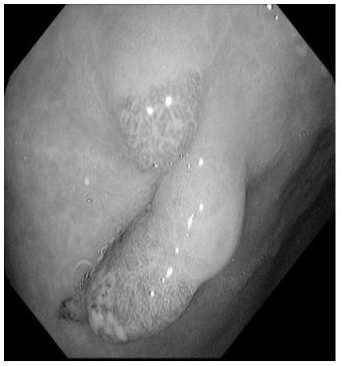
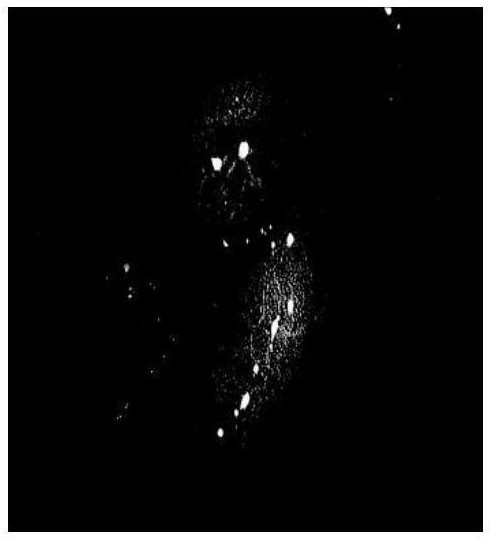
[0059] In the process of obtaining the repaired image, the specular reflection needs to be removed. Before removing the specular reflection, the highlight point detection should be carried out. The method used to detect the highlight point is a threshold algorithm, and the image blocks larger than a certain threshold are divided according to the size of the highlight point. In addition, ...

Embodiment 2

[0103] First, the image to be repaired is image-enhanced, and then repaired using traditional algorithms. Four-fifths of the repaired polyp data set are selected as the training set and the corresponding mask images marked by experts are put into three types. The commonly used medical polyp segmentation network is trained. Then put the remaining one-fifth of the restored image data set as a test set into the Unet, Unet++, and PraNet networks that have been trained for comparison. The segmentation results of the Unet network are compared, for example Figure 5(a)-Figure 5(b) shown; comparison of segmentation results of Unet++ network Figure 6(a)-Figure 6(b) shown; the comparison of the segmentation results of the PraNet network is as follows Figure 7(a)-Figure 7(b) shown; by comparing the segmentation results when there are highlights and the segmentation results when there are no highlights, the improvement of the segmentation effect can be observed; Unet is a network propo...


PUM


Abstract

The invention discloses a medical image segmentation method based on highlight spot removal, and relates to the related technical field of image recognition, and the method specifically comprises the steps: obtaining a to-be-restored image; performing image enhancement processing on the to-be-restored image; repairing the enhanced to-be-repaired image to obtain a repaired image; training an image segmentation model by using a generative adversarial network based on the repaired image; performing image segmentation on the repaired image by using the image segmentation model; according to the method, most of the bright spots in the medical image can be restored, and the texture can be kept not lost, so that the image is more suitable for aesthetic appreciation of human eyes, and the visual experience is not influenced by the high bright spots.

Description

technical field [0001] The invention relates to the technical field of image recognition, and more particularly to a medical image segmentation method based on highlight removal. Background technique [0002] With the development of science and technology, minimally invasive medical procedures have become more and more common as medical means; medical image segmentation is a complex and critical step in the field of medical image processing and analysis. Part of it is segmented, and relevant features are extracted to provide a reliable basis for clinical diagnosis and treatment and pathological research, and assist doctors to make more accurate diagnosis. Since the FCN fully convolutional network was proposed, research in the field of medical image segmentation has been widely replaced by deep learning methods. However, due to the complexity of the medical image itself and the specular reflection phenomenon of the endoscopic image, the endoscopic image has highlights. Doct...

Claims


Application Information

Patent Timeline
Patent Type & Authority Applications(China)
IPC IPC(8): G06T7/10G06T7/13G06T7/136G06T5/00G06T5/30G06N3/04G06N3/08
CPCG06T7/10G06T7/136G06T7/13G06T5/30G06N3/084G06T2207/20081G06T2207/20084G06N3/045G06T5/77
Inventor 徐超钱凯李正平
Owner ANHUI UNIVERSITY
Learn More