Intracardiac multi-mode ultrasonic imaging method, device and system

An ultrasonic imaging method and ultrasonic imaging technology, applied in the field of medical imaging, can solve the problems of high cost, precise positioning of difficult lesion areas, and high equipment requirements, and achieve the effect of facilitating diagnosis and being beneficial to clinical diagnosis.

Pending Publication Date: 2022-07-15
SHENZHEN INSIGHTSONICS CO LTD
View PDF0 Cites 3 Cited by
  • Summary
  • Abstract
  • Description
  • Claims
  • Application Information

AI Technical Summary

Problems solved by technology

[0006] For the guidance and monitoring of myocardial radiofrequency ablation, although the real-time two-dimensional ultrasound B images used by ICE can provide information on cardiac cavity structure and tissue morphology, it is difficult to accurately locate the lesion area and the actual ablated area based on this
Although the use of magnetic resonance temperature imaging can realize quasi-real-time monitoring of the ablation process, it requires high equipment, complicated operation and high cost
For the measurement of mechanical parameters of in vivo cardiac tissue, due to the position of the heart, it is difficult for existing in vitro ultrasound methods to flexibly and accurately perform large-scale and multi-dimensional mechanical measurements of in vivo cardiac tissue

Method used


Image

  • Intracardiac multi-mode ultrasonic imaging method, device and system
  • Intracardiac multi-mode ultrasonic imaging method, device and system
  • Intracardiac multi-mode ultrasonic imaging method, device and system

Examples


Embodiment 1

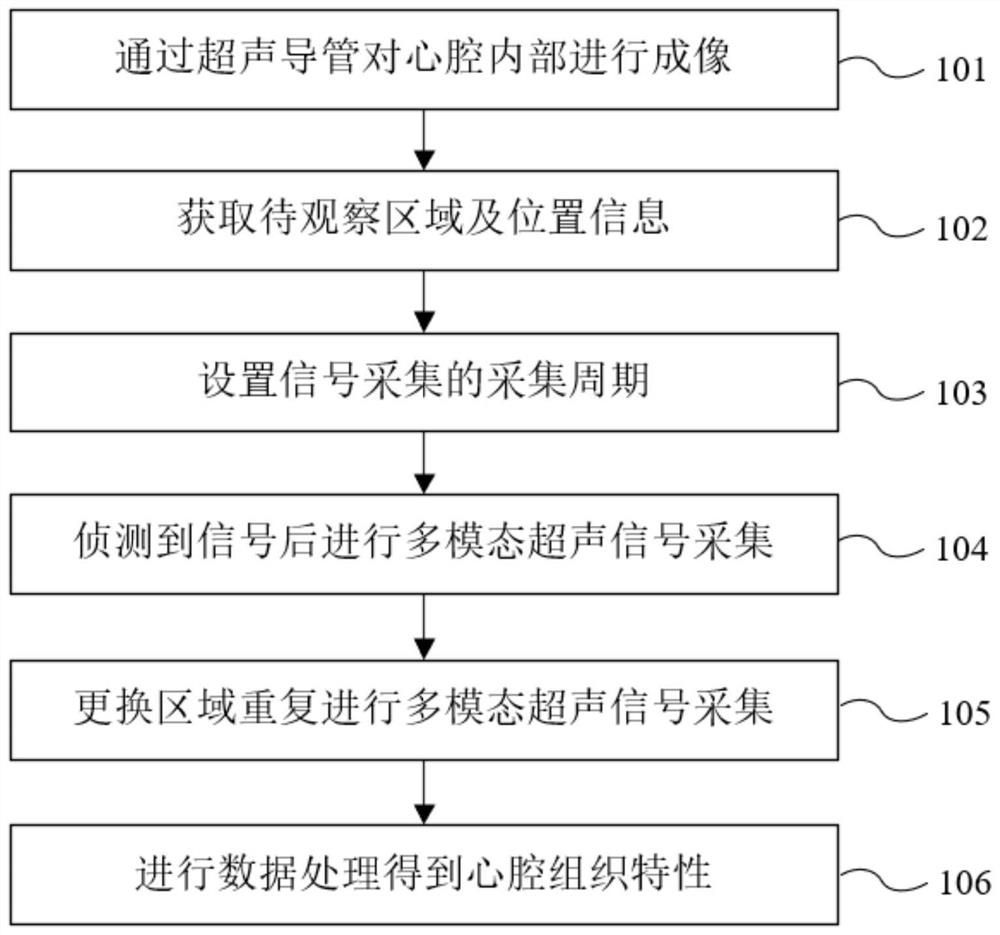
[0059] This embodiment proposes an intracardiac cavity multimodal ultrasound imaging method, which is used to measure the time-varying three-dimensional cardiac cavity structure, myocardial tissue displacement, strain, elasticity and other characteristics from the interior of the cardiac cavity, so as to realize the intracardiac cavity tissue characteristics acquisition. The flow chart of intracardiac multimodal ultrasound imaging method is attached to the instruction manual figure 1 As shown, the specific scheme is as follows:

[0060] An intracardiac multimodal ultrasound imaging method, comprising the following steps:

[0061] 101. Insert the ultrasound imaging catheter connected with the probe into the cardiac cavity along the blood vessel, and perform real-time B-mode imaging inside the cardiac cavity;

[0062] 102. Adjust the position of the probe to obtain the area to be observed and the position information corresponding to the probe;

[0063] 103. Set the acquisiti...

Embodiment 2

[0107] This embodiment provides an intracardiac multimodal ultrasound imaging device, which systematizes the method of Embodiment 1. The schematic diagram of the system structure is shown in the appendix of the description. image 3 As shown, the specific scheme is as follows:

[0108] An intracardiac multimodal ultrasound imaging device, comprising the following:

[0109] Detection unit A1: used to insert the ultrasound imaging catheter connected with the probe into the cardiac cavity along the blood vessel, and perform real-time B-mode imaging inside the cardiac cavity;

[0110] Area acquisition unit A2: used to adjust the position of the probe, and obtain the area to be observed and the position information corresponding to the probe;

[0111] Period setting unit A3: used to set the acquisition period of the preset multimodal ultrasound signal acquisition, taking a certain moment in a heartbeat cycle as the acquisition start time, and the acquisition start time shows a spe...

Embodiment 3

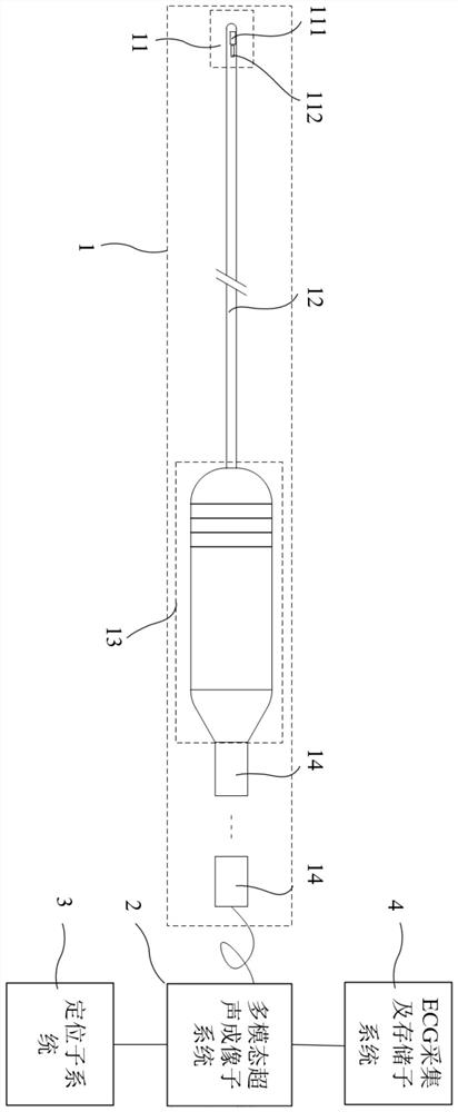
[0119] This embodiment provides an intracardiac multimodal ultrasound imaging system, which is used to implement the intracardiac multimodal ultrasound imaging method of Embodiment 1. A schematic structural diagram of the system is shown in the appendix of the description. figure 2 As shown, the specific scheme is as follows:

[0120] An intracardiac multimodal ultrasound imaging system, comprising the following:

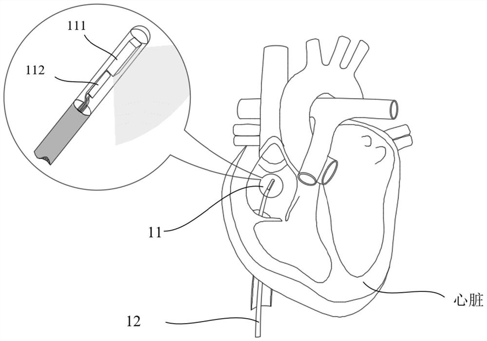
[0121] The ultrasonic imaging catheter subsystem 1 includes a probe 11 and an ultrasonic imaging catheter 12 connected to each other, an ultrasonic transducer 111 and a positioning device 112 are arranged on the probe 11, and are used for inserting the ultrasonic imaging catheter 12 together with the probe 11 into a cardiac cavity along a blood vessel, Real-time B-mode imaging is performed on the interior of the cardiac cavity by transmitting and receiving signals from the ultrasonic transducer 111 ; wherein, the probe 11 is also provided with a positioning device ...


PUM


Abstract

The invention provides an intracardiac multi-mode ultrasonic imaging method, device and system, and the method comprises the steps: enabling an ultrasonic imaging catheter to be inserted into a cardiac cavity along a blood vessel, and carrying out the real-time B-mode imaging of the interior of the cardiac cavity; adjusting the position of the probe; setting an acquisition period of multi-modal ultrasonic signal acquisition, and adjusting the position information to perform multi-modal ultrasonic signal acquisition on the plurality of to-be-observed areas; and performing data processing on the ultrasonic signals under each piece of position information, and obtaining the cardiac cavity tissue characteristics of each to-be-observed area in the acquisition period in combination with a time sequence. According to the scheme, four-dimensional multi-mode imaging and parameter measurement can be carried out on the heart tissue, related information such as a three-dimensional heart cavity structure, myocardial tissue displacement, strain and elasticity at different moments of a heartbeat cycle is measured from the interior of a heart cavity, the heart cavity tissue characteristics of the heart tissue are obtained, and multi-dimensional mechanical parameter measurement is achieved.

Description

technical field [0001] The present invention relates to the field of medical imaging, and in particular, to an intracardiac multimodal ultrasound imaging method, device and system. Background technique [0002] Intracardiac echocardiography (ICE) is a technique for real-time imaging of intracardiac structures by placing a catheter containing an ultrasound probe into the heart chamber through a blood vessel. Compared with conventional in vitro cardiac ultrasound imaging, ICE can help doctors to see the structure of the heart cavity more clearly and intuitively, and is often used to guide myocardial radiofrequency ablation (RFA) to treat atrial fibrillation and other arrhythmias. . [0003] However, the real-time 2D ultrasound B image used by ICE can only provide information on the structure and tissue morphology of the cardiac cavity, and it is difficult to accurately control the position and size of the ablation site and the process of ablation for guiding myocardial radiof...

Claims


Application Information

Patent Timeline
Patent Type & Authority Applications(China)
IPC IPC(8): A61B8/12A61B8/08A61B8/00
CPCA61B8/12A61B8/0883A61B8/4411A61B8/52A61B8/5207A61B8/5246A61B8/485A61B8/4254
Inventor 王亚华陈明心
Owner SHENZHEN INSIGHTSONICS CO LTD
Why Eureka
Try Eureka