Enhanced CT image rectal cancer staging auxiliary diagnosis system based on deep learning

A CT image and deep learning technology, applied in the field of medical image processing, can solve the problem of difficult to distinguish cancerous areas and their stages, and achieve the effect of improving segmentation accuracy and accuracy.

Pending Publication Date: 2022-07-22
ANHUI MEDICAL UNIV +3
View PDF1 Cites 5 Cited by
  • Summary
  • Abstract
  • Description
  • Claims
  • Application Information

AI Technical Summary

Problems solved by technology

[0004] In view of the defects in the above-mentioned prior art, the purpose of the present invention is to provide an enhanced CT image rectal cancer staging auxiliary diagnosis system based on deep learning, which is also an auxiliary diagnostic device, for the complex structure of rectal enhanced CT image data, and it is difficult to distinguish cancer. The problem of region and its staging, research on enhanced CT image rectal cancer data labeling and data set construction, rectal cancer lesion region discrimination based on self-attention deep learning model, metastatic lymph node recognition based on sequence adaptive feature fusion, design and implementation of rectal cancer Staging intelligent auxiliary diagnosis system, and conduct clinical application experiment verification to improve the accuracy and efficiency of comprehensive data collection before rectal cancer surgery

Method used


Image

  • Enhanced CT image rectal cancer staging auxiliary diagnosis system based on deep learning
  • Enhanced CT image rectal cancer staging auxiliary diagnosis system based on deep learning
  • Enhanced CT image rectal cancer staging auxiliary diagnosis system based on deep learning

Examples


Embodiment Construction

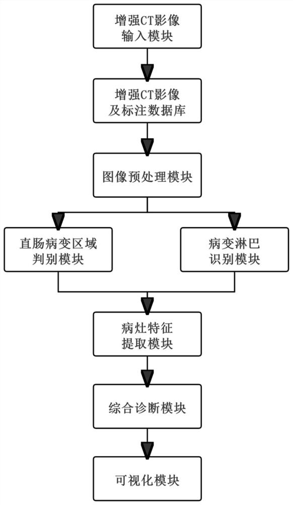
[0059] refer to figure 1 The present invention provides an enhanced CT image rectal cancer staging auxiliary diagnosis system based on deep learning, including an enhanced CT image input module, an enhanced CT image and an annotation database, an image preprocessing module, a rectal lesion area discrimination module, and a lesion lymph identification module , Lesion feature extraction module, comprehensive diagnosis module, visualization module.

[0060] The execution of the device includes the following steps:

[0061] S1. Construct an enhanced CT rectal cancer image dataset and annotated database;

[0062] S2. Perform format conversion and image noise reduction processing on the enhanced CT image;

[0063] S3. The rectal lesion area discrimination module is based on the self-attention deep learning model to discriminate whether the CT image contains a suspected rectal tumor (lesion) shape area, and segment the lesion area;

[0064] S4. The diseased lymph node identificati...


PUM


Abstract

The invention relates to an enhanced CT image rectal cancer staging auxiliary diagnosis system based on deep learning. The system comprises an enhanced CT image input module, an enhanced CT image and annotation database, an image preprocessing module, a rectal lesion area discrimination module, a lesion lymph recognition module, a focus feature extraction module, a comprehensive diagnosis module and a visualization module. Aiming at the problems that a rectum enhanced CT image is complex in data structure and difficult to distinguish cancerous areas and stages thereof, the rectum enhanced CT image rectal cancer data labeling and data set construction, rectal cancer lesion area discrimination based on a self-attention deep learning model and metastatic lymph node identification based on sequence adaptive feature fusion are researched. A rectal cancer staging intelligent auxiliary diagnosis system is designed and realized, and clinical application experiment verification is carried out, so that comprehensive data acquisition accuracy and efficiency before a rectal cancer operation are improved.

Description

technical field [0001] The invention belongs to the technical field of medical image processing, and in particular relates to an enhanced CT image rectal cancer staging auxiliary diagnosis system based on deep learning. Background technique [0002] Rectal cancer is the most common malignant tumor of the digestive system, which generally refers to the cancer from the dentate line to the junction of the rectosigmoid colon, and is one of the most common malignant tumors of the digestive tract. Rectal cancer is located in a low position and is easily diagnosed by digital rectal examination and colonoscopy. However, due to its location deep into the pelvis, complex anatomical relationships, and limitations of inspection methods, it is difficult to achieve accurate preoperative staging diagnosis. The staging and diagnosis of rectal cancer is crucial for accurate and individualized treatment decisions, and is a prerequisite for ensuring a good prognosis for patients. X-ray, MRI ...

Claims


Application Information

Patent Timeline
Patent Type & Authority Applications(China)
IPC IPC(8): G06T7/00G06T5/00G06T7/11G16H30/20G16H50/20G06N3/08G06K9/62G06V10/774G06V10/82
CPCG06T7/0012G06T7/11G06T5/002G06N3/08G16H50/20G16H30/20G06T2207/10081G06T2207/30028G06T2207/30096G06F18/214
Inventor 邹兵兵万寿红王万勤邱晨阳张翰韬刘宏武毕军焱
Owner ANHUI MEDICAL UNIV
Why Eureka
Try Eureka